 Movie
Movie Controller

[English] 日本語
 Yorodumi
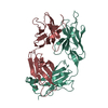
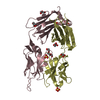
- PDB-6zvf: Crystal structure of the recombinant Fab fragment derived from th... -
+ Open data

- Basic information
| Entry | Database: PDB / ID: 6zvf | ||||||
|---|---|---|---|---|---|---|---|
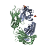
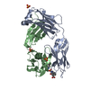
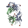
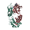
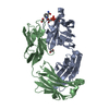
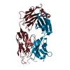
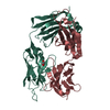
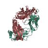
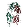
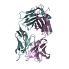
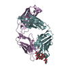
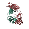
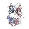
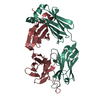
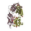
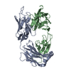
| Title | Crystal structure of the recombinant Fab fragment derived from the hybridoma M3/38 in complex with a human Galectin-3 peptide | ||||||
 Components Components |
| ||||||
 Keywords Keywords |  IMMUNE SYSTEM / ANTIBODY / FAB FRAGMENT / PEPTIDE EPITOPE / PROTEIN ENGINEERING IMMUNE SYSTEM / ANTIBODY / FAB FRAGMENT / PEPTIDE EPITOPE / PROTEIN ENGINEERING | ||||||
| Function / homology |  Function and homology information Function and homology informationnegative regulation of protein tyrosine phosphatase activity / negative regulation of immunological synapse formation / negative regulation of T cell activation via T cell receptor contact with antigen bound to MHC molecule on antigen presenting cell / RUNX2 regulates genes involved in differentiation of myeloid cells / disaccharide binding / regulation of T cell apoptotic process / mononuclear cell migration / IgE binding / negative regulation of endocytosis / positive regulation of mononuclear cell migration ...negative regulation of protein tyrosine phosphatase activity / negative regulation of immunological synapse formation / negative regulation of T cell activation via T cell receptor contact with antigen bound to MHC molecule on antigen presenting cell / RUNX2 regulates genes involved in differentiation of myeloid cells / disaccharide binding / regulation of T cell apoptotic process / mononuclear cell migration / IgE binding / negative regulation of endocytosis / positive regulation of mononuclear cell migration / eosinophil chemotaxis / regulation of extrinsic apoptotic signaling pathway via death domain receptors / RUNX1 regulates transcription of genes involved in differentiation of myeloid cells / protein phosphatase inhibitor activity / negative regulation of T cell receptor signaling pathway / positive chemotaxis / regulation of T cell proliferation / positive regulation of calcium ion import / macrophage chemotaxis / chemoattractant activity / monocyte chemotaxis / Advanced glycosylation endproduct receptor signaling / ficolin-1-rich granule membrane / immunological synapse / laminin binding / epithelial cell differentiation / neutrophil chemotaxis / RNA splicing / secretory granule membrane / molecular condensate scaffold activity / negative regulation of extrinsic apoptotic signaling pathway / positive regulation of protein localization to plasma membrane / spliceosomal complex / positive regulation of protein-containing complex assembly / mRNA processing / carbohydrate binding / protein phosphatase binding / collagen-containing extracellular matrix / mitochondrial inner membrane / innate immune response / Neutrophil degranulation / cell surface / extracellular space / RNA binding / extracellular exosome / extracellular region / nucleoplasm / membrane / nucleus / plasma membrane / cytosol / cytoplasmSimilarity search - Function | ||||||
| Biological species |  Rattus norvegicus (Norway rat) Rattus norvegicus (Norway rat) Homo sapiens (human) Homo sapiens (human) | ||||||
| Method | X-RAY DIFFRACTION / SYNCHROTRON / MOLECULAR REPLACEMENT / molecular replacement / Resolution: 1.9 Å | ||||||
 Authors Authors | Skerra, A. / Eichinger, A. | ||||||
| Funding support |  Germany, 1items Germany, 1items
| ||||||
 Citation Citation | Journal: Sci Rep / Year: 2021 Title: Effective rational humanization of a PASylated anti-galectin-3 Fab for the sensitive PET imaging of thyroid cancer in vivo. Authors: Peplau, E. / De Rose, F. / Eichinger, A. / Reder, S. / Mittelhauser, M. / Scafetta, G. / Schwaiger, M. / Weber, W.A. / Bartolazzi, A. / D'Alessandria, C. / Skerra, A. | ||||||
| History |
|
- Structure visualization
| Structure viewer | Molecule: MolmilJmol/JSmol |
|---|
- Downloads & links
-Download
| PDBx/mmCIF format | 6zvf.cif.gz | 184.3 KB | Display | PDBx/mmCIF format |
|---|---|---|---|---|
| PDB format | pdb6zvf.ent.gz | 145.7 KB | Display | PDB format |
| PDBx/mmJSON format | 6zvf.json.gz | Tree view | PDBx/mmJSON format | |
| Others |  Other downloads Other downloads |
-Validation report
| Arichive directory | https://data.pdbj.org/pub/pdb/validation_reports/zv/6zvfftp://data.pdbj.org/pub/pdb/validation_reports/zv/6zvf | HTTPS FTP |
|---|
-Related structure data
| Related structure data |  3o2vS S: Starting model for refinement |
|---|---|
| Similar structure data |


-Links
 PDBj
PDBj


- Assembly
| Deposited unit | 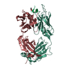
| ||||||||
|---|---|---|---|---|---|---|---|---|---|
| 1 |
| ||||||||
| Unit cell |
|
-Components
| #1: Antibody | Mass: 23927.803 Da / Num. of mol.: 1 Source method: isolated from a genetically manipulated source Details: Both immunoglobulin chains (L, H) are linked by a disulfide bond (CYS219L to CYS221H). Source: (gene. exp.) Rattus norvegicus (Norway rat) / Production host:  Escherichia coli (E. coli) Escherichia coli (E. coli) |
|---|---|
| #2: Antibody | Mass: 24392.615 Da / Num. of mol.: 1 Source method: isolated from a genetically manipulated source Details: Residues 222 - 227 represent an affinity tag (His6-tag). Both immunoglobulin chains (L, H) are linked by a disulfide bond (CYS219L to CYS221H). Source: (gene. exp.) Rattus norvegicus (Norway rat) / Production host: Escherichia coli (E. coli) |
| #3: Protein/peptide | / Gal-3 / 35 kDa lectin / Carbohydrate-binding protein 35 / CBP 35 / Galactose-specific lectin 3 / ...Gal-3 / 35 kDa lectin / Carbohydrate-binding protein 35 / CBP 35 / Galactose-specific lectin 3 / Galactoside-binding protein / GALBP / IgE-binding protein / L-31 / Laminin-binding protein / Lectin L-29 / Mac-2 antigen Mass: 882.959 Da / Num. of mol.: 1 / Source method: obtained synthetically Details: The peptide QAPPGAYPG carries an acetyl (ACE) group at the N-terminus. Source: (synth.) Homo sapiens (human) / References: UniProt: P17931 |
| #4: Water | ChemComp-HOH / Water Mass: 18.015 Da / Num. of mol.: 201 / Source method: isolated from a natural source / Formula: H2O Mass: 18.015 Da / Num. of mol.: 201 / Source method: isolated from a natural source / Formula: H2O |
| Has ligand of interest | N |
-Experimental details
-Experiment
| Experiment | Method: X-RAY DIFFRACTION / Number of used crystals: 1 |
|---|
- Sample preparation
| Crystal | Density Matthews: 2.31 Å3/Da / Density % sol: 46.68 % |
|---|---|
| Crystal grow | Temperature: 293 K / Method: vapor diffusion, hanging drop / pH: 7 / Details: polyethylene glycol 4000 |
-Data collection
| Diffraction | Mean temperature: 100 K / Serial crystal experiment: N | |||||||||||||||||||||||||||||||||||||||||||||||||||||||||||||||||||||||||||||||||||||||||||||||||||||||||||||||||||||||||
|---|---|---|---|---|---|---|---|---|---|---|---|---|---|---|---|---|---|---|---|---|---|---|---|---|---|---|---|---|---|---|---|---|---|---|---|---|---|---|---|---|---|---|---|---|---|---|---|---|---|---|---|---|---|---|---|---|---|---|---|---|---|---|---|---|---|---|---|---|---|---|---|---|---|---|---|---|---|---|---|---|---|---|---|---|---|---|---|---|---|---|---|---|---|---|---|---|---|---|---|---|---|---|---|---|---|---|---|---|---|---|---|---|---|---|---|---|---|---|---|---|---|---|
| Diffraction source | Source: SYNCHROTRON / Site: BESSY / Beamline: 14.1 / Wavelength: 0.9184 Å | |||||||||||||||||||||||||||||||||||||||||||||||||||||||||||||||||||||||||||||||||||||||||||||||||||||||||||||||||||||||||
| Detector | Type: DECTRIS PILATUS 6M / Detector: PIXEL / Date: Mar 22, 2018 / Details: Si mirror | |||||||||||||||||||||||||||||||||||||||||||||||||||||||||||||||||||||||||||||||||||||||||||||||||||||||||||||||||||||||||
| Radiation | Monochromator: Si 111 double-crystal / Protocol: SINGLE WAVELENGTH / Monochromatic (M) / Laue (L): M / Scattering type: x-ray | |||||||||||||||||||||||||||||||||||||||||||||||||||||||||||||||||||||||||||||||||||||||||||||||||||||||||||||||||||||||||
| Radiation wavelength | Wavelength: 0.9184 Å / Relative weight: 1 | |||||||||||||||||||||||||||||||||||||||||||||||||||||||||||||||||||||||||||||||||||||||||||||||||||||||||||||||||||||||||
| Reflection | Resolution: 1.661→78.719 Å / Num. all: 34354 / Num. obs: 34354 / % possible obs: 97 % / Redundancy: 5.7 % / Rpim(I) all: 0.039 / Rrim(I) all: 0.096 / Rsym value: 0.088 / Net I/av σ(I): 5.7 / Net I/σ(I): 10.9 / Num. measured all: 197033 | |||||||||||||||||||||||||||||||||||||||||||||||||||||||||||||||||||||||||||||||||||||||||||||||||||||||||||||||||||||||||
| Reflection shell | Diffraction-ID: 1
|
-Phasing
| Phasing | Method: molecular replacement | |||||||||
|---|---|---|---|---|---|---|---|---|---|---|
| Phasing MR | Model details: Phaser MODE: MR_AUTO
|
- Processing
| Software |
| ||||||||||||||||||||||||||||||||||||||||||||||||||||||||||||||||||||||||||||||||||||||||||||||||||||
|---|---|---|---|---|---|---|---|---|---|---|---|---|---|---|---|---|---|---|---|---|---|---|---|---|---|---|---|---|---|---|---|---|---|---|---|---|---|---|---|---|---|---|---|---|---|---|---|---|---|---|---|---|---|---|---|---|---|---|---|---|---|---|---|---|---|---|---|---|---|---|---|---|---|---|---|---|---|---|---|---|---|---|---|---|---|---|---|---|---|---|---|---|---|---|---|---|---|---|---|---|---|
| Refinement | Method to determine structure: MOLECULAR REPLACEMENT Starting model: 3O2V Resolution: 1.9→50.98 Å / Cor.coef. Fo:Fc: 0.943 / Cor.coef. Fo:Fc free: 0.921 / SU B: 8.385 / SU ML: 0.12 / Cross valid method: THROUGHOUT / σ(F): 0 / ESU R: 0.177 / ESU R Free: 0.159 / Stereochemistry target values: MAXIMUM LIKELIHOOD Details: HYDROGENS HAVE BEEN ADDED IN THE RIDING POSITIONS U VALUES : WITH TLS ADDED
| ||||||||||||||||||||||||||||||||||||||||||||||||||||||||||||||||||||||||||||||||||||||||||||||||||||
| Solvent computation | Ion probe radii: 0.8 Å / Shrinkage radii: 0.8 Å / VDW probe radii: 1.2 Å / Solvent model: MASK | ||||||||||||||||||||||||||||||||||||||||||||||||||||||||||||||||||||||||||||||||||||||||||||||||||||
| Displacement parameters | Biso max: 88.63 Å2 / Biso mean: 25.385 Å2 / Biso min: 12.98 Å2
| ||||||||||||||||||||||||||||||||||||||||||||||||||||||||||||||||||||||||||||||||||||||||||||||||||||
| Refinement step | Cycle: final / Resolution: 1.9→50.98 Å
| ||||||||||||||||||||||||||||||||||||||||||||||||||||||||||||||||||||||||||||||||||||||||||||||||||||
| Refine LS restraints |
| ||||||||||||||||||||||||||||||||||||||||||||||||||||||||||||||||||||||||||||||||||||||||||||||||||||
| LS refinement shell | Resolution: 1.9→1.949 Å / Rfactor Rfree error: 0 / Total num. of bins used: 20
| ||||||||||||||||||||||||||||||||||||||||||||||||||||||||||||||||||||||||||||||||||||||||||||||||||||
| Refinement TLS params. | Method: refined / Refine-ID: X-RAY DIFFRACTION
| ||||||||||||||||||||||||||||||||||||||||||||||||||||||||||||||||||||||||||||||||||||||||||||||||||||
| Refinement TLS group |
|
