 Movie
Movie Controller

[English] 日本語
 Yorodumi
- PDB-6zue: Crystal structure of human DDB1 bound to human DCAF1 (amino acid ... -
+ Open data

- Basic information
| Entry | Database: PDB / ID: 6zue | ||||||
|---|---|---|---|---|---|---|---|
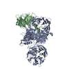
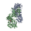
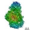
| Title | Crystal structure of human DDB1 bound to human DCAF1 (amino acid residues 1046-1396) | ||||||
 Components Components |
| ||||||
 Keywords Keywords |  PROTEIN BINDING / Cullin-RING E3 ubiquitin ligase / Cullin 4 / ubiquitylation / DNA repair PROTEIN BINDING / Cullin-RING E3 ubiquitin ligase / Cullin 4 / ubiquitylation / DNA repair | ||||||
| Function / homology |  Function and homology information Function and homology informationhistone H2AT120 kinase activity / cell competition in a multicellular organism / positive regulation by virus of viral protein levels in host cell / V(D)J recombination / epigenetic programming in the zygotic pronuclei / spindle assembly involved in female meiosis / Cul4-RING E3 ubiquitin ligase complex / UV-damage excision repair / biological process involved in interaction with symbiont / WD40-repeat domain binding ...histone H2AT120 kinase activity / cell competition in a multicellular organism / positive regulation by virus of viral protein levels in host cell / V(D)J recombination / epigenetic programming in the zygotic pronuclei / spindle assembly involved in female meiosis / Cul4-RING E3 ubiquitin ligase complex / UV-damage excision repair / biological process involved in interaction with symbiont / WD40-repeat domain binding / regulation of mitotic cell cycle phase transition / Cul4A-RING E3 ubiquitin ligase complex / Cul4B-RING E3 ubiquitin ligase complex / ubiquitin ligase complex scaffold activity / negative regulation of reproductive process / negative regulation of developmental process / cullin family protein binding / ubiquitin-like ligase-substrate adaptor activity / viral release from host cell / ectopic germ cell programmed cell death / positive regulation of viral genome replication / positive regulation of gluconeogenesis / post-translational protein modification / B cell differentiation / proteasomal protein catabolic process / nuclear estrogen receptor binding / nucleotide-excision repair / Recognition of DNA damage by PCNA-containing replication complex / DNA Damage Recognition in GG-NER / regulation of circadian rhythm / Dual Incision in GG-NER / Transcription-Coupled Nucleotide Excision Repair (TC-NER) / Formation of TC-NER Pre-Incision Complex / fibrillar center / Wnt signaling pathway / Formation of Incision Complex in GG-NER / Dual incision in TC-NER / Gap-filling DNA repair synthesis and ligation in TC-NER / positive regulation of protein catabolic process / rhythmic process / cellular response to UV / protein-macromolecule adaptor activity / Antigen processing: Ubiquitination & Proteasome degradation / site of double-strand break / Neddylation / ubiquitin-dependent protein catabolic process / proteasome-mediated ubiquitin-dependent protein catabolic process / chromosome, telomeric region / damaged DNA binding / protein ubiquitination / non-specific serine/threonine protein kinase / phosphorylation / protein serine kinase activity / DNA repair / apoptotic process / DNA damage response / protein-containing complex binding / nucleolus / negative regulation of apoptotic process / negative regulation of transcription by RNA polymerase II / protein-containing complex / DNA binding / extracellular space / extracellular exosome / nucleoplasm / ATP binding / nucleus / cytoplasmSimilarity search - Function | ||||||
| Biological species |  Homo sapiens (human) Homo sapiens (human) | ||||||
| Method | X-RAY DIFFRACTION / SYNCHROTRON / MOLECULAR REPLACEMENT / Resolution: 3.094 Å | ||||||
 Authors Authors | Schwefel, D. / Taylor, I.A. | ||||||
| Funding support |  Germany, 1items Germany, 1items
| ||||||
 Citation Citation | Journal: Plos Pathog. / Year: 2021 Title: Structural insights into Cullin4-RING ubiquitin ligase remodelling by Vpr from simian immunodeficiency viruses. Authors: Banchenko, S. / Krupp, F. / Gotthold, C. / Burger, J. / Graziadei, A. / O'Reilly, F.J. / Sinn, L. / Ruda, O. / Rappsilber, J. / Spahn, C.M.T. / Mielke, T. / Taylor, I.A. / Schwefel, D. | ||||||
| History |
|
- Structure visualization
| Structure viewer | Molecule: MolmilJmol/JSmol |
|---|
- Downloads & links
-Download
| PDBx/mmCIF format | 6zue.cif.gz | 580.7 KB | Display | PDBx/mmCIF format |
|---|---|---|---|---|
| PDB format | pdb6zue.ent.gz | 478.7 KB | Display | PDB format |
| PDBx/mmJSON format | 6zue.json.gz | Tree view | PDBx/mmJSON format | |
| Others |  Other downloads Other downloads |
-Validation report
| Arichive directory | https://data.pdbj.org/pub/pdb/validation_reports/zu/6zueftp://data.pdbj.org/pub/pdb/validation_reports/zu/6zue | HTTPS FTP |
|---|
-Related structure data
| Related structure data |  6zx9C 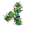 3e0cS S: Starting model for refinement C: citing same article ( |
|---|---|
| Similar structure data |


-Links
 PDBj
PDBj


- Assembly
| Deposited unit | 
| ||||||||
|---|---|---|---|---|---|---|---|---|---|
| 1 |
| ||||||||
| Unit cell |
|
-Components
| #1: Protein | Mass: 127241.594 Da / Num. of mol.: 1 Source method: isolated from a genetically manipulated source Source: (gene. exp.) Homo sapiens (human) / Gene: DDB1, XAP1 / Cell line (production host): Sf9 / Production host:   Spodoptera frugiperda (fall armyworm) / References: UniProt: Q16531 Spodoptera frugiperda (fall armyworm) / References: UniProt: Q16531 |
|---|---|
| #2: Protein | Mass: 42502.770 Da / Num. of mol.: 1 Source method: isolated from a genetically manipulated source Source: (gene. exp.) Homo sapiens (human) / Gene: DCAF1, KIAA0800, RIP, VPRBP / Cell line (production host): Sf9 / Production host: Spodoptera frugiperda (fall armyworm)References: UniProt: Q9Y4B6, non-specific serine/threonine protein kinase |
| #3: Water | ChemComp-HOH / Water Mass: 18.015 Da / Num. of mol.: 10 / Source method: isolated from a natural source / Formula: H2O Mass: 18.015 Da / Num. of mol.: 10 / Source method: isolated from a natural source / Formula: H2O |
-Experimental details
-Experiment
| Experiment | Method: X-RAY DIFFRACTION / Number of used crystals: 1 |
|---|
- Sample preparation
| Crystal | Density Matthews: 2.96 Å3/Da / Density % sol: 58.49 % |
|---|---|
| Crystal grow | Temperature: 291 K / Method: vapor diffusion, hanging drop / pH: 5.5 / Details: 100 mM Tri-Na citrate pH 5.5 18% PEG 1000 |
-Data collection
| Diffraction | Mean temperature: 100 K / Serial crystal experiment: N |
|---|---|
| Diffraction source | Source: SYNCHROTRON / Site: Diamond  / Beamline: I04 / Wavelength: 0.92819 Å / Beamline: I04 / Wavelength: 0.92819 Å |
| Detector | Type: DECTRIS PILATUS3 S 6M / Detector: PIXEL / Date: Jan 17, 2016 |
| Radiation | Protocol: SINGLE WAVELENGTH / Monochromatic (M) / Laue (L): M / Scattering type: x-ray |
| Radiation wavelength | Wavelength: 0.92819 Å / Relative weight: 1 |
| Reflection | Resolution: 3.09→50 Å / Num. obs: 36027 / % possible obs: 96.8 % / Redundancy: 4.82 % / CC1/2: 0.999 / Net I/σ(I): 12.98 |
| Reflection shell | Resolution: 3.09→3.28 Å / Num. unique obs: 5769 / CC1/2: 0.853 |
- Processing
| Software |
| ||||||||||||||||||||||||||||||||||||||||||||||||||||||||||||||||||||||||||||||||||||||||||||||||||||||||||||||||||||||||||||||||||||||||||||||||||||||||||||||||||||||||||||||||||||||||||||||||||||||||
|---|---|---|---|---|---|---|---|---|---|---|---|---|---|---|---|---|---|---|---|---|---|---|---|---|---|---|---|---|---|---|---|---|---|---|---|---|---|---|---|---|---|---|---|---|---|---|---|---|---|---|---|---|---|---|---|---|---|---|---|---|---|---|---|---|---|---|---|---|---|---|---|---|---|---|---|---|---|---|---|---|---|---|---|---|---|---|---|---|---|---|---|---|---|---|---|---|---|---|---|---|---|---|---|---|---|---|---|---|---|---|---|---|---|---|---|---|---|---|---|---|---|---|---|---|---|---|---|---|---|---|---|---|---|---|---|---|---|---|---|---|---|---|---|---|---|---|---|---|---|---|---|---|---|---|---|---|---|---|---|---|---|---|---|---|---|---|---|---|---|---|---|---|---|---|---|---|---|---|---|---|---|---|---|---|---|---|---|---|---|---|---|---|---|---|---|---|---|---|---|---|---|
| Refinement | Method to determine structure: MOLECULAR REPLACEMENT Starting model: 3e0c Resolution: 3.094→48.634 Å / SU ML: 0.41 / Cross valid method: THROUGHOUT / σ(F): 1.35 / Phase error: 33.12 / Stereochemistry target values: ML
| ||||||||||||||||||||||||||||||||||||||||||||||||||||||||||||||||||||||||||||||||||||||||||||||||||||||||||||||||||||||||||||||||||||||||||||||||||||||||||||||||||||||||||||||||||||||||||||||||||||||||
| Solvent computation | Shrinkage radii: 0.9 Å / VDW probe radii: 1.11 Å / Solvent model: FLAT BULK SOLVENT MODEL | ||||||||||||||||||||||||||||||||||||||||||||||||||||||||||||||||||||||||||||||||||||||||||||||||||||||||||||||||||||||||||||||||||||||||||||||||||||||||||||||||||||||||||||||||||||||||||||||||||||||||
| Displacement parameters | Biso max: 274.9 Å2 / Biso mean: 113.7834 Å2 / Biso min: 45.66 Å2 | ||||||||||||||||||||||||||||||||||||||||||||||||||||||||||||||||||||||||||||||||||||||||||||||||||||||||||||||||||||||||||||||||||||||||||||||||||||||||||||||||||||||||||||||||||||||||||||||||||||||||
| Refinement step | Cycle: final / Resolution: 3.094→48.634 Å
| ||||||||||||||||||||||||||||||||||||||||||||||||||||||||||||||||||||||||||||||||||||||||||||||||||||||||||||||||||||||||||||||||||||||||||||||||||||||||||||||||||||||||||||||||||||||||||||||||||||||||
| Refine LS restraints |
| ||||||||||||||||||||||||||||||||||||||||||||||||||||||||||||||||||||||||||||||||||||||||||||||||||||||||||||||||||||||||||||||||||||||||||||||||||||||||||||||||||||||||||||||||||||||||||||||||||||||||
| LS refinement shell | Refine-ID: X-RAY DIFFRACTION / Rfactor Rfree error: 0
| ||||||||||||||||||||||||||||||||||||||||||||||||||||||||||||||||||||||||||||||||||||||||||||||||||||||||||||||||||||||||||||||||||||||||||||||||||||||||||||||||||||||||||||||||||||||||||||||||||||||||
| Refinement TLS params. | Method: refined / Refine-ID: X-RAY DIFFRACTION
| ||||||||||||||||||||||||||||||||||||||||||||||||||||||||||||||||||||||||||||||||||||||||||||||||||||||||||||||||||||||||||||||||||||||||||||||||||||||||||||||||||||||||||||||||||||||||||||||||||||||||
| Refinement TLS group |
|
