 Movie
Movie Controller


+ Open data

- Basic information
| Entry | Database: PDB / ID: 6zr7 | ||||||
|---|---|---|---|---|---|---|---|
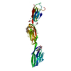
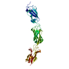
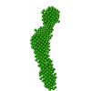
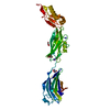
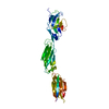
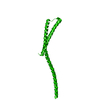
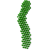
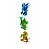
| Title | X-ray structure of human Dscam Ig7-Ig9 | ||||||
 Components Components | Down syndrome cell adhesion molecule | ||||||
 Keywords Keywords |  CELL ADHESION / neuronal self-avoidance / N-glycosylation CELL ADHESION / neuronal self-avoidance / N-glycosylation | ||||||
| Function / homology |  Function and homology information Function and homology informationpost-embryonic retina morphogenesis in camera-type eye / camera-type eye photoreceptor cell differentiation / netrin receptor binding / retina layer formation / dendrite self-avoidance / cell-cell adhesion mediator activity / positive regulation of axon extension involved in axon guidance / negative regulation of cell adhesion / dendrite morphogenesis / DSCAM interactions ...post-embryonic retina morphogenesis in camera-type eye / camera-type eye photoreceptor cell differentiation / netrin receptor binding / retina layer formation / dendrite self-avoidance / cell-cell adhesion mediator activity / positive regulation of axon extension involved in axon guidance / negative regulation of cell adhesion / dendrite morphogenesis / DSCAM interactions / homophilic cell adhesion via plasma membrane adhesion molecules / plasma membrane => GO:0005886 / positive regulation of phosphorylation / synapse assembly / protein tyrosine kinase binding / locomotory behavior / axon guidance / nervous system development / growth cone / cell adhesion / axon / neuronal cell body / synapse / dendrite / extracellular region / membrane / plasma membraneSimilarity search - Function | ||||||
| Biological species |  Homo sapiens (human) Homo sapiens (human) | ||||||
| Method | X-RAY DIFFRACTION / SYNCHROTRON / MOLECULAR REPLACEMENT / Resolution: 1.85 Å | ||||||
 Authors Authors | Kozak, S. / Bento, I. / Meijers, R. | ||||||
 Citation Citation | Journal: Acta Crystallogr D Struct Biol / Year: 2020 Title: Homogeneously N-glycosylated proteins derived from the GlycoDelete HEK293 cell line enable diffraction-quality crystallogenesis. Authors: Kozak, S. / Bloch, Y. / De Munck, S. / Mikula, A. / Bento, I. / Savvides, S.N. / Meijers, R. | ||||||
| History |
|
- Structure visualization
| Structure viewer | Molecule: MolmilJmol/JSmol |
|---|
- Downloads & links
-Download
| PDBx/mmCIF format | 6zr7.cif.gz | 86.6 KB | Display | PDBx/mmCIF format |
|---|---|---|---|---|
| PDB format | pdb6zr7.ent.gz | Display | PDB format | |
| PDBx/mmJSON format | 6zr7.json.gz | Tree view | PDBx/mmJSON format | |
| Others |  Other downloads Other downloads |
-Validation report
| Arichive directory | https://data.pdbj.org/pub/pdb/validation_reports/zr/6zr7ftp://data.pdbj.org/pub/pdb/validation_reports/zr/6zr7 | HTTPS FTP |
|---|
-Related structure data
| Related structure data |  6sffC  4wvrS S: Starting model for refinement C: citing same article ( |
|---|---|
| Similar structure data |


-Links
 PDBj
PDBj


- Assembly
| Deposited unit | 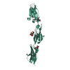
| ||||||||
|---|---|---|---|---|---|---|---|---|---|
| 1 |
| ||||||||
| Unit cell |
|
-Components
-Protein , 1 types, 1 molecules AAA
| #1: Protein | Mass: 34254.012 Da / Num. of mol.: 1 Source method: isolated from a genetically manipulated source Source: (gene. exp.) Homo sapiens (human) / Gene: DSCAM / Production host: Homo sapiens (human) / References: UniProt: O60469 |
|---|
-Sugars , 3 types, 4 molecules 
| #2: Polysaccharide | N-acetyl-alpha-neuraminic acid-(2-3)-beta-D-galactopyranose-(1-4)-2-acetamido-2-deoxy-beta-D-glucopyranose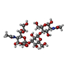  , Oligosaccharide / Class: Substrate analog / Mass: 674.604 Da / Num. of mol.: 1 , Oligosaccharide / Class: Substrate analog / Mass: 674.604 Da / Num. of mol.: 1Source method: isolated from a genetically manipulated source Details: oligosaccharide / References: 3'-sialyl-N-acetyllactosamine | ||
|---|---|---|---|
| #3: Polysaccharide | / Mass: 383.349 Da / Num. of mol.: 2 / Source method: obtained synthetically| #6: Sugar | ChemComp-NAG / | N-Acetylglucosamine Type: D-saccharide, beta linking / Mass: 221.208 Da / Num. of mol.: 1 Type: D-saccharide, beta linking / Mass: 221.208 Da / Num. of mol.: 1Source method: isolated from a genetically manipulated source Formula: C8H15NO6 |
-Non-polymers , 3 types, 168 molecules 


| #4: Chemical | ChemComp-GOL / Glycerol Mass: 92.094 Da / Num. of mol.: 1 / Source method: obtained synthetically / Formula: C3H8O3 Mass: 92.094 Da / Num. of mol.: 1 / Source method: obtained synthetically / Formula: C3H8O3 |
|---|---|
| #5: Chemical | ChemComp-CA /  Mass: 40.078 Da / Num. of mol.: 1 Mass: 40.078 Da / Num. of mol.: 1Source method: isolated from a genetically manipulated source Formula: Ca |
| #7: Water | ChemComp-HOH / WaterMass: 18.015 Da / Num. of mol.: 166 / Source method: isolated from a natural source / Formula: H2O |
-Details
| Has ligand of interest | N |
|---|
-Experimental details
-Experiment
| Experiment | Method: X-RAY DIFFRACTION / Number of used crystals: 1 |
|---|
- Sample preparation
| Crystal | Density Matthews: 3.48 Å3/Da / Density % sol: 64.72 % |
|---|---|
| Crystal grow | Temperature: 292 K / Method: vapor diffusion / pH: 7.5 / Details: Glycerol, calcium acetate, PEG 8000, HEPES |
-Data collection
| Diffraction | Mean temperature: 100 K / Serial crystal experiment: N |
|---|---|
| Diffraction source | Source: SYNCHROTRON / Site: PETRA III, EMBL c/o DESY  / Beamline: P13 (MX1) / Wavelength: 0.9763 Å / Beamline: P13 (MX1) / Wavelength: 0.9763 Å |
| Detector | Type: DECTRIS PILATUS 6M / Detector: PIXEL / Date: Dec 7, 2017 |
| Radiation | Monochromator: Double Crystal Monochromator FMB Oxford / Protocol: SINGLE WAVELENGTH / Monochromatic (M) / Laue (L): M / Scattering type: x-ray |
| Radiation wavelength | Wavelength: 0.9763 Å / Relative weight: 1 |
| Reflection | Resolution: 1.85→84.81 Å / Num. obs: 39662 / % possible obs: 99 % / Redundancy: 3.8 % / Biso Wilson estimate: 36.88 Å2 / CC1/2: 1 / Rmerge(I) obs: 0.044 / Rpim(I) all: 0.026 / Rrim(I) all: 0.051 / Net I/σ(I): 12.8 |
| Reflection shell | Resolution: 1.85→1.89 Å / Rmerge(I) obs: 0.874 / Num. unique obs: 2418 / CC1/2: 0.7 / Rpim(I) all: 0.501 / Rrim(I) all: 1.011 / % possible all: 97.4 |
- Processing
| Software |
| |||||||||||||||||||||||||||||||||||||||||||||||||||||||||||||||||||||||||||||||||||||||||||||||||||||||||||||||||||||||||||||||||||||||||||||||||||||||||||||||||||||||||||||||||||||||||||||||||||||||||||||||||||||||||||||||||||||||
|---|---|---|---|---|---|---|---|---|---|---|---|---|---|---|---|---|---|---|---|---|---|---|---|---|---|---|---|---|---|---|---|---|---|---|---|---|---|---|---|---|---|---|---|---|---|---|---|---|---|---|---|---|---|---|---|---|---|---|---|---|---|---|---|---|---|---|---|---|---|---|---|---|---|---|---|---|---|---|---|---|---|---|---|---|---|---|---|---|---|---|---|---|---|---|---|---|---|---|---|---|---|---|---|---|---|---|---|---|---|---|---|---|---|---|---|---|---|---|---|---|---|---|---|---|---|---|---|---|---|---|---|---|---|---|---|---|---|---|---|---|---|---|---|---|---|---|---|---|---|---|---|---|---|---|---|---|---|---|---|---|---|---|---|---|---|---|---|---|---|---|---|---|---|---|---|---|---|---|---|---|---|---|---|---|---|---|---|---|---|---|---|---|---|---|---|---|---|---|---|---|---|---|---|---|---|---|---|---|---|---|---|---|---|---|---|---|---|---|---|---|---|---|---|---|---|---|---|---|---|---|---|---|
| Refinement | Method to determine structure: MOLECULAR REPLACEMENT Starting model: 4wvr Resolution: 1.85→84.808 Å / Cor.coef. Fo:Fc: 0.962 / Cor.coef. Fo:Fc free: 0.955 / WRfactor Rfree: 0.243 / WRfactor Rwork: 0.217 / SU B: 3.016 / SU ML: 0.088 / Average fsc free: 0.8942 / Average fsc work: 0.9078 / Cross valid method: FREE R-VALUE / ESU R: 0.113 / ESU R Free: 0.112 Details: Hydrogens have been added in their riding positions
| |||||||||||||||||||||||||||||||||||||||||||||||||||||||||||||||||||||||||||||||||||||||||||||||||||||||||||||||||||||||||||||||||||||||||||||||||||||||||||||||||||||||||||||||||||||||||||||||||||||||||||||||||||||||||||||||||||||||
| Solvent computation | Ion probe radii: 0.8 Å / Shrinkage radii: 0.8 Å / VDW probe radii: 1.2 Å / Solvent model: MASK BULK SOLVENT | |||||||||||||||||||||||||||||||||||||||||||||||||||||||||||||||||||||||||||||||||||||||||||||||||||||||||||||||||||||||||||||||||||||||||||||||||||||||||||||||||||||||||||||||||||||||||||||||||||||||||||||||||||||||||||||||||||||||
| Displacement parameters | Biso mean: 56.811 Å2
| |||||||||||||||||||||||||||||||||||||||||||||||||||||||||||||||||||||||||||||||||||||||||||||||||||||||||||||||||||||||||||||||||||||||||||||||||||||||||||||||||||||||||||||||||||||||||||||||||||||||||||||||||||||||||||||||||||||||
| Refinement step | Cycle: LAST / Resolution: 1.85→84.808 Å
| |||||||||||||||||||||||||||||||||||||||||||||||||||||||||||||||||||||||||||||||||||||||||||||||||||||||||||||||||||||||||||||||||||||||||||||||||||||||||||||||||||||||||||||||||||||||||||||||||||||||||||||||||||||||||||||||||||||||
| Refine LS restraints |
| |||||||||||||||||||||||||||||||||||||||||||||||||||||||||||||||||||||||||||||||||||||||||||||||||||||||||||||||||||||||||||||||||||||||||||||||||||||||||||||||||||||||||||||||||||||||||||||||||||||||||||||||||||||||||||||||||||||||
| LS refinement shell | Refine-ID: X-RAY DIFFRACTION / Total num. of bins used: 20
|
