 Movie
Movie Controller

[English] 日本語
 Yorodumi
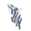
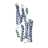
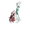
- PDB-6zh1: Crystal structure of complex between FH19-20 and FhbA protein fro... -
+ Open data

- Basic information
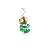
| Entry | Database: PDB / ID: 6zh1 | |||||||||
|---|---|---|---|---|---|---|---|---|---|---|
| Title | Crystal structure of complex between FH19-20 and FhbA protein from Borrelia hermsii | |||||||||
 Components Components |
| |||||||||
 Keywords Keywords |  IMMUNE SYSTEM / Innate immunity / complement / relapsing fever / Lyme disease IMMUNE SYSTEM / Innate immunity / complement / relapsing fever / Lyme disease | |||||||||
| Function / homology |  Function and homology informationregulation of complement activation, alternative pathway / symbiont cell surface / complement component C3b binding / regulation of complement-dependent cytotoxicity / regulation of complement activation / heparan sulfate proteoglycan binding / serine-type endopeptidase complex / complement activation, alternative pathway / complement activation / Regulation of Complement cascade ...regulation of complement activation, alternative pathway / symbiont cell surface / complement component C3b binding / regulation of complement-dependent cytotoxicity / regulation of complement activation / heparan sulfate proteoglycan binding / serine-type endopeptidase complex / complement activation, alternative pathway / complement activation / Regulation of Complement cascade / heparin binding / blood microparticle / proteolysis / extracellular space / extracellular exosome / extracellular region / identical protein binding Function and homology informationregulation of complement activation, alternative pathway / symbiont cell surface / complement component C3b binding / regulation of complement-dependent cytotoxicity / regulation of complement activation / heparan sulfate proteoglycan binding / serine-type endopeptidase complex / complement activation, alternative pathway / complement activation / Regulation of Complement cascade ...regulation of complement activation, alternative pathway / symbiont cell surface / complement component C3b binding / regulation of complement-dependent cytotoxicity / regulation of complement activation / heparan sulfate proteoglycan binding / serine-type endopeptidase complex / complement activation, alternative pathway / complement activation / Regulation of Complement cascade / heparin binding / blood microparticle / proteolysis / extracellular space / extracellular exosome / extracellular region / identical protein bindingSimilarity search - Function | |||||||||
| Biological species |  Borrelia hermsii YOR (bacteria) Borrelia hermsii YOR (bacteria) Homo sapiens (human) Homo sapiens (human) | |||||||||
| Method | X-RAY DIFFRACTION / SYNCHROTRON / MOLECULAR REPLACEMENT / molecular replacement / Resolution: 2.2 Å | |||||||||
 Authors Authors | Kogan, K. / Kotila, T. / Meri, T. / Goldman, A. | |||||||||
| Funding support |  Finland, Finland,  United Kingdom, 2items United Kingdom, 2items
| |||||||||
 Citation Citation | Journal: Plos Pathog. / Year: 2022 Title: Mechanism of Borrelia immune evasion by FhbA-related proteins. Authors: Kogan, K. / Haapasalo, K. / Kotila, T. / Moore, R. / Lappalainen, P. / Goldman, A. / Meri, T. | |||||||||
| History |
|
- Structure visualization
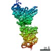
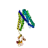
| Structure viewer | Molecule: MolmilJmol/JSmol |
|---|
- Downloads & links
-Download
| PDBx/mmCIF format | 6zh1.cif.gz | 139.6 KB | Display | PDBx/mmCIF format |
|---|---|---|---|---|
| PDB format | pdb6zh1.ent.gz | 107.5 KB | Display | PDB format |
| PDBx/mmJSON format | 6zh1.json.gz | Tree view | PDBx/mmJSON format | |
| Others |  Other downloads Other downloads |
-Validation report
| Arichive directory | https://data.pdbj.org/pub/pdb/validation_reports/zh/6zh1ftp://data.pdbj.org/pub/pdb/validation_reports/zh/6zh1 | HTTPS FTP |
|---|
-Related structure data
| Related structure data | 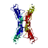 2g7iS S: Starting model for refinement |
|---|---|
| Similar structure data |
-Links
 PDBj
PDBj

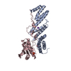
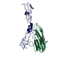
- Assembly
| Deposited unit | 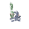
| ||||||||
|---|---|---|---|---|---|---|---|---|---|
| 1 |
| ||||||||
| Unit cell |
|
-Components
| #1: Protein | Mass: 22148.938 Da / Num. of mol.: 1 Source method: isolated from a genetically manipulated source Details: Factor H-binding protein A / Source: (gene. exp.) Borrelia hermsii YOR (bacteria) / Gene: BHY_1174 / Production host: Escherichia coli BL21(DE3) (bacteria) / References: UniProt: W5SB08 | ||||||
|---|---|---|---|---|---|---|---|
| #2: Protein | Factor H / H factor 1 Mass: 15084.103 Da / Num. of mol.: 1 Source method: isolated from a genetically manipulated source Source: (gene. exp.) Homo sapiens (human) / Gene: CFH, HF, HF1, HF2 / Production host:  Komagataella pastoris (fungus) / References: UniProt: P08603 Komagataella pastoris (fungus) / References: UniProt: P08603 | ||||||
| #3: Chemical | ChemComp-EDO / Ethylene glycol  Mass: 62.068 Da / Num. of mol.: 4 / Source method: obtained synthetically / Formula: C2H6O2 Mass: 62.068 Da / Num. of mol.: 4 / Source method: obtained synthetically / Formula: C2H6O2#4: Chemical | ChemComp-SO4 / Sulfate  Mass: 96.063 Da / Num. of mol.: 5 / Source method: obtained synthetically / Formula: SO4 Mass: 96.063 Da / Num. of mol.: 5 / Source method: obtained synthetically / Formula: SO4#5: Water | ChemComp-HOH / | Water Mass: 18.015 Da / Num. of mol.: 154 / Source method: isolated from a natural source / Formula: H2O Mass: 18.015 Da / Num. of mol.: 154 / Source method: isolated from a natural source / Formula: H2OHas ligand of interest | N | |
-Experimental details
-Experiment
| Experiment | Method: X-RAY DIFFRACTION / Number of used crystals: 1 |
|---|
- Sample preparation
| Crystal | Density Matthews: 2.85 Å3/Da / Density % sol: 56.91 % |
|---|---|
| Crystal grow | Temperature: 293.15 K / Method: vapor diffusion, hanging drop / pH: 6.7 Details: 0.1 M MES pH6.7, 0.2 M Ammonium Sulfate, 20% (v/v) PEG 4000 |
-Data collection
| Diffraction | Mean temperature: 100 K / Serial crystal experiment: N |
|---|---|
| Diffraction source | Source: SYNCHROTRON / Site: ESRF  / Beamline: ID23-2 / Wavelength: 0.8729 Å / Beamline: ID23-2 / Wavelength: 0.8729 Å |
| Detector | Type: DECTRIS PILATUS3 2M / Detector: PIXEL / Date: Oct 29, 2014 |
| Radiation | Protocol: SINGLE WAVELENGTH / Monochromatic (M) / Laue (L): M / Scattering type: x-ray |
| Radiation wavelength | Wavelength: 0.8729 Å / Relative weight: 1 |
| Reflection | Resolution: 2.2→37.828 Å / Num. obs: 20180 / % possible obs: 100 % / Redundancy: 13.1 % / Biso Wilson estimate: 49.19 Å2 / CC1/2: 0.999 / Rmerge(I) obs: 0.166 / Rpim(I) all: 0.047 / Rrim(I) all: 0.172 / Net I/av σ(I): 12.531 / Net I/σ(I): 12.531 |
| Reflection shell | Resolution: 2.2→2.237 Å / Redundancy: 13.78 % / Rmerge(I) obs: 2.436 / Mean I/σ(I) obs: 1 / Num. unique obs: 993 / CC1/2: 0.513 / Rpim(I) all: 0.673 / Rrim(I) all: 2.529 / % possible all: 99 |
-Phasing
| Phasing | Method: molecular replacement |
|---|
- Processing
| Software |
| ||||||||||||||||||||||||||||||||||||||||||||||||||||||||||||||||||||||||||||||||||||||||||||||||||||||||||||
|---|---|---|---|---|---|---|---|---|---|---|---|---|---|---|---|---|---|---|---|---|---|---|---|---|---|---|---|---|---|---|---|---|---|---|---|---|---|---|---|---|---|---|---|---|---|---|---|---|---|---|---|---|---|---|---|---|---|---|---|---|---|---|---|---|---|---|---|---|---|---|---|---|---|---|---|---|---|---|---|---|---|---|---|---|---|---|---|---|---|---|---|---|---|---|---|---|---|---|---|---|---|---|---|---|---|---|---|---|---|
| Refinement | Method to determine structure: MOLECULAR REPLACEMENT Starting model: 2G7I Resolution: 2.2→22.88 Å / Cor.coef. Fo:Fc: 0.939 / Cor.coef. Fo:Fc free: 0.915 / SU R Cruickshank DPI: 0.211 / Cross valid method: THROUGHOUT / σ(F): 0 / SU R Blow DPI: 0.227 / SU Rfree Blow DPI: 0.192 / SU Rfree Cruickshank DPI: 0.187
| ||||||||||||||||||||||||||||||||||||||||||||||||||||||||||||||||||||||||||||||||||||||||||||||||||||||||||||
| Displacement parameters | Biso max: 160.42 Å2 / Biso mean: 59.9 Å2 / Biso min: 27.31 Å2
| ||||||||||||||||||||||||||||||||||||||||||||||||||||||||||||||||||||||||||||||||||||||||||||||||||||||||||||
| Refine analyze | Luzzati coordinate error obs: 0.32 Å | ||||||||||||||||||||||||||||||||||||||||||||||||||||||||||||||||||||||||||||||||||||||||||||||||||||||||||||
| Refinement step | Cycle: final / Resolution: 2.2→22.88 Å
| ||||||||||||||||||||||||||||||||||||||||||||||||||||||||||||||||||||||||||||||||||||||||||||||||||||||||||||
| Refine LS restraints |
| ||||||||||||||||||||||||||||||||||||||||||||||||||||||||||||||||||||||||||||||||||||||||||||||||||||||||||||
| LS refinement shell | Resolution: 2.2→2.32 Å / Rfactor Rfree error: 0 / Total num. of bins used: 10
| ||||||||||||||||||||||||||||||||||||||||||||||||||||||||||||||||||||||||||||||||||||||||||||||||||||||||||||
| Refinement TLS params. | Method: refined / Refine-ID: X-RAY DIFFRACTION
| ||||||||||||||||||||||||||||||||||||||||||||||||||||||||||||||||||||||||||||||||||||||||||||||||||||||||||||
| Refinement TLS group |
|
