 Movie
Movie Controller

[English] 日本語
 Yorodumi
- PDB-2g7i: Structure of Human Complement Factor H Carboxyl Terminal Domains ... -
+ Open data

- Basic information
| Entry | Database: PDB / ID: 2g7i | ||||||
|---|---|---|---|---|---|---|---|
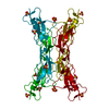
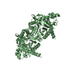
| Title | Structure of Human Complement Factor H Carboxyl Terminal Domains 19-20: a Basis for Atypical Hemolytic Uremic Syndrome | ||||||
 Components Components | Complement factor H Factor H Factor H | ||||||
 Keywords Keywords | IMMUNE SYSTEM / Sushi (CCP/SCR) domain / Complement / Regulator / Factor H / Beta-1H Globulin / Atypical Hemolytic Uremic Syndrome / Heparin Binding | ||||||
| Function / homology |  Function and homology informationregulation of complement activation, alternative pathway / symbiont cell surface / complement component C3b binding / regulation of complement-dependent cytotoxicity / regulation of complement activation / heparan sulfate proteoglycan binding / serine-type endopeptidase complex / complement activation, alternative pathway / complement activation / Regulation of Complement cascade ...regulation of complement activation, alternative pathway / symbiont cell surface / complement component C3b binding / regulation of complement-dependent cytotoxicity / regulation of complement activation / heparan sulfate proteoglycan binding / serine-type endopeptidase complex / complement activation, alternative pathway / complement activation / Regulation of Complement cascade / heparin binding / blood microparticle / proteolysis / extracellular space / extracellular exosome / extracellular region / identical protein binding Function and homology informationregulation of complement activation, alternative pathway / symbiont cell surface / complement component C3b binding / regulation of complement-dependent cytotoxicity / regulation of complement activation / heparan sulfate proteoglycan binding / serine-type endopeptidase complex / complement activation, alternative pathway / complement activation / Regulation of Complement cascade ...regulation of complement activation, alternative pathway / symbiont cell surface / complement component C3b binding / regulation of complement-dependent cytotoxicity / regulation of complement activation / heparan sulfate proteoglycan binding / serine-type endopeptidase complex / complement activation, alternative pathway / complement activation / Regulation of Complement cascade / heparin binding / blood microparticle / proteolysis / extracellular space / extracellular exosome / extracellular region / identical protein bindingSimilarity search - Function | ||||||
| Biological species |  Homo sapiens (human) Homo sapiens (human) | ||||||
| Method | X-RAY DIFFRACTION / SYNCHROTRON / SAD / Resolution: 1.75 Å | ||||||
 Authors Authors | Jaakola, V.-P. / Jokiranta, T.S. / Goldman, A. | ||||||
 Citation Citation | Journal: Embo J. / Year: 2006 Title: Structure of complement factor H carboxyl-terminus reveals molecular basis of atypical haemolytic uremic syndrome. Authors: Jokiranta, T.S. / Jaakola, V.-P. / Lehtinen, M.J. / Parepalo, M. / Meri, S. / Goldman, A. | ||||||
| History |
|
- Structure visualization
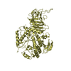
| Structure viewer | Molecule: MolmilJmol/JSmol |
|---|
- Downloads & links
-Download
| PDBx/mmCIF format | 2g7i.cif.gz | 38.3 KB | Display | PDBx/mmCIF format |
|---|---|---|---|---|
| PDB format | pdb2g7i.ent.gz | 29.8 KB | Display | PDB format |
| PDBx/mmJSON format | 2g7i.json.gz | Tree view | PDBx/mmJSON format | |
| Others |  Other downloads Other downloads |
-Validation report
| Arichive directory | https://data.pdbj.org/pub/pdb/validation_reports/g7/2g7iftp://data.pdbj.org/pub/pdb/validation_reports/g7/2g7i | HTTPS FTP |
|---|
-Related structure data
| Similar structure data |
|---|

-Links
 PDBj
PDBj

- Assembly
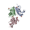
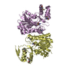
| Deposited unit | 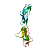
| ||||||||
|---|---|---|---|---|---|---|---|---|---|
| 1 | 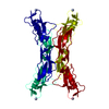
| ||||||||
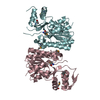
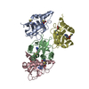
| Unit cell |
| ||||||||
| Details | Crystal packing of FH19-20 in the asymmetric unit reveals tightly packed tetrameric assembly of domains of FH19-20; may have biological relevance |
-Components
| #1: Protein | Factor H / H factor 1 Mass: 14297.312 Da / Num. of mol.: 1 / Fragment: C-terminal Domains 19-20 Source method: isolated from a genetically manipulated source Source: (gene. exp.) Homo sapiens (human) / Gene: CFH, HF, HF1 / Plasmid: pPICZ B expression vector / Production host:  Pichia pastoris (fungus) / References: UniProt: P08603 Pichia pastoris (fungus) / References: UniProt: P08603 |
|---|---|
| #2: Water | ChemComp-HOH / Water Mass: 18.015 Da / Num. of mol.: 177 / Source method: isolated from a natural source / Formula: H2O Mass: 18.015 Da / Num. of mol.: 177 / Source method: isolated from a natural source / Formula: H2O |
-Experimental details
-Experiment
| Experiment | Method: X-RAY DIFFRACTION / Number of used crystals: 2 |
|---|
- Sample preparation
| Crystal | Density Matthews: 4.05 Å3/Da / Density % sol: 69.61 % |
|---|---|
| Crystal grow | Temperature: 298 K / Method: vapor diffusion, sitting drop / pH: 8.5 Details: Before crystallization the protein was concentrated to 10 mg/ml and dialysed into 20 mM Tris, 50 mM NaCl, pH 7.0. The protein was crystallized in sitting drops by mixing 1 ul of protein ...Details: Before crystallization the protein was concentrated to 10 mg/ml and dialysed into 20 mM Tris, 50 mM NaCl, pH 7.0. The protein was crystallized in sitting drops by mixing 1 ul of protein solution at 10 mg/ml with 1 ul of reservoir solution of 2.2 M (NH4)2SO4, 0.1 M Tris-HCl, pH 8.5, VAPOR DIFFUSION, SITTING DROP, temperature 298.0K |
-Data collection
| Diffraction |
| ||||||||||||||||||
|---|---|---|---|---|---|---|---|---|---|---|---|---|---|---|---|---|---|---|---|
| Diffraction source |
| ||||||||||||||||||
| Detector |
| ||||||||||||||||||
| Radiation | Protocol: SINGLE WAVELENGTH / Monochromatic (M) / Laue (L): M / Scattering type: x-ray | ||||||||||||||||||
| Radiation wavelength |
| ||||||||||||||||||
| Reflection | Resolution: 1.75→14.78 Å / Num. all: 22700 / Num. obs: 22700 / % possible obs: 99.93 % / Observed criterion σ(F): 0 / Observed criterion σ(I): 0 / Redundancy: 6.9 % / Rsym value: 0.059 | ||||||||||||||||||
| Reflection shell | Resolution: 1.75→1.795 Å / Redundancy: 5.1 % / Mean I/σ(I) obs: 3.7 / Rsym value: 0.441 / % possible all: 99.9 |

- Processing
| Software |
| |||||||||||||||||||||||||||||||||||||||||||||||||||||||||||||||||||||||||||||||||||||||||||||||||||||||||||||||||||||||||||||
|---|---|---|---|---|---|---|---|---|---|---|---|---|---|---|---|---|---|---|---|---|---|---|---|---|---|---|---|---|---|---|---|---|---|---|---|---|---|---|---|---|---|---|---|---|---|---|---|---|---|---|---|---|---|---|---|---|---|---|---|---|---|---|---|---|---|---|---|---|---|---|---|---|---|---|---|---|---|---|---|---|---|---|---|---|---|---|---|---|---|---|---|---|---|---|---|---|---|---|---|---|---|---|---|---|---|---|---|---|---|---|---|---|---|---|---|---|---|---|---|---|---|---|---|---|---|---|
| Refinement | Method to determine structure: SAD / Resolution: 1.75→14.78 Å / Cor.coef. Fo:Fc: 0.942 / Cor.coef. Fo:Fc free: 0.935 / SU B: 1.631 / SU ML: 0.055 / Cross valid method: THROUGHOUT / σ(F): 0 / ESU R: 0.093 / ESU R Free: 0.09 / Stereochemistry target values: MAXIMUM LIKELIHOOD / Details: HYDROGENS HAVE BEEN ADDED IN THE RIDING POSITIONS
| |||||||||||||||||||||||||||||||||||||||||||||||||||||||||||||||||||||||||||||||||||||||||||||||||||||||||||||||||||||||||||||
| Solvent computation | Ion probe radii: 0.8 Å / Shrinkage radii: 0.8 Å / VDW probe radii: 1.2 Å / Solvent model: MASK | |||||||||||||||||||||||||||||||||||||||||||||||||||||||||||||||||||||||||||||||||||||||||||||||||||||||||||||||||||||||||||||
| Displacement parameters | Biso mean: 23.593 Å2
| |||||||||||||||||||||||||||||||||||||||||||||||||||||||||||||||||||||||||||||||||||||||||||||||||||||||||||||||||||||||||||||
| Refinement step | Cycle: LAST / Resolution: 1.75→14.78 Å
| |||||||||||||||||||||||||||||||||||||||||||||||||||||||||||||||||||||||||||||||||||||||||||||||||||||||||||||||||||||||||||||
| Refine LS restraints |
| |||||||||||||||||||||||||||||||||||||||||||||||||||||||||||||||||||||||||||||||||||||||||||||||||||||||||||||||||||||||||||||
| LS refinement shell | Resolution: 1.75→1.795 Å / Total num. of bins used: 20
|
