 Movie
Movie Controller

[English] 日本語
 Yorodumi
- PDB-6zab: Structure of the transcriptional repressor Atu1419 (VanR) from ag... -
+ Open data

- Basic information
| Entry | Database: PDB / ID: 6zab | ||||||
|---|---|---|---|---|---|---|---|
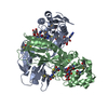
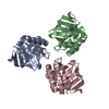
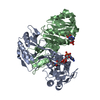
| Title | Structure of the transcriptional repressor Atu1419 (VanR) from agrobacterium fabrum in complex a palindromic DNA (P6422 space group) | ||||||
 Components Components |
| ||||||
 Keywords Keywords |  TRANSCRIPTION / repressor TRANSCRIPTION / repressor | ||||||
| Function / homology |  Function and homology information Function and homology information | ||||||
| Biological species |  Agrobacterium fabrum (bacteria)Agrobacterium fabrum str. C58 (bacteria) Agrobacterium fabrum (bacteria)Agrobacterium fabrum str. C58 (bacteria) | ||||||
| Method | X-RAY DIFFRACTION / SYNCHROTRON / MOLECULAR REPLACEMENT / molecular replacement / Resolution: 2.8 Å | ||||||
 Authors Authors | Morera, S. / Naretto, A. / Vigouroux, A. / Legrand, P. | ||||||
 Citation Citation | Journal: Nucleic Acids Res. / Year: 2021 Title: Characterization of the first tetrameric transcription factor of the GntR superfamily with allosteric regulation from the bacterial pathogen Agrobacterium fabrum. Authors: Vigouroux, A. / Meyer, T. / Naretto, A. / Legrand, P. / Aumont-Nicaise, M. / Di Cicco, A. / Renoud, S. / Dore, J. / Levy, D. / Vial, L. / Lavire, C. / Morera, S. | ||||||
| History |
|
- Structure visualization
| Structure viewer | Molecule: MolmilJmol/JSmol |
|---|
- Downloads & links
-Download
| PDBx/mmCIF format | 6zab.cif.gz | 125.1 KB | Display | PDBx/mmCIF format |
|---|---|---|---|---|
| PDB format | pdb6zab.ent.gz | 94.1 KB | Display | PDB format |
| PDBx/mmJSON format | 6zab.json.gz | Tree view | PDBx/mmJSON format | |
| Others |  Other downloads Other downloads |
-Validation report
| Arichive directory | https://data.pdbj.org/pub/pdb/validation_reports/za/6zabftp://data.pdbj.org/pub/pdb/validation_reports/za/6zab | HTTPS FTP |
|---|
-Related structure data
| Related structure data | 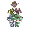 6z74SC 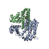 6za0C  6za3C 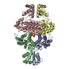 6za7C S: Starting model for refinement C: citing same article ( |
|---|---|
| Similar structure data |


-Links
 PDBj
PDBj- Assembly
| Deposited unit | 
| ||||||||
|---|---|---|---|---|---|---|---|---|---|
| 1 | 
| ||||||||
| Unit cell |
|
-Components
-Protein / DNA chain , 2 types, 2 molecules AB
| #1: Protein | Transcriptional regulation Mass: 27407.182 Da / Num. of mol.: 1 Source method: isolated from a genetically manipulated source Source: (gene. exp.) Agrobacterium fabrum (strain C58 / ATCC 33970) (bacteria)Strain: C58 / ATCC 33970 / Gene: Atu1419 / Production host: Escherichia coli (E. coli) / References: UniProt: A9CJ36 |
|---|---|
| #2: DNA chain | Mass: 3043.029 Da / Num. of mol.: 1 Source method: isolated from a genetically manipulated source Source: (gene. exp.) Agrobacterium fabrum str. C58 (bacteria)Production host: synthetic construct (others) |
-Non-polymers , 5 types, 27 molecules 


| #3: Chemical | ChemComp-ZN /  Mass: 65.409 Da / Num. of mol.: 1 / Source method: obtained synthetically / Formula: Zn / Feature type: SUBJECT OF INVESTIGATION Mass: 65.409 Da / Num. of mol.: 1 / Source method: obtained synthetically / Formula: Zn / Feature type: SUBJECT OF INVESTIGATION |
|---|---|
| #4: Chemical | ChemComp-CIT / Citric acid Mass: 192.124 Da / Num. of mol.: 1 / Source method: obtained synthetically / Formula: C6H8O7 Mass: 192.124 Da / Num. of mol.: 1 / Source method: obtained synthetically / Formula: C6H8O7 |
| #5: Chemical | ChemComp-PEG / Diethylene glycol Mass: 106.120 Da / Num. of mol.: 1 / Source method: obtained synthetically / Formula: C4H10O3 Mass: 106.120 Da / Num. of mol.: 1 / Source method: obtained synthetically / Formula: C4H10O3 |
| #6: Chemical | ChemComp-EDO / Ethylene glycol Mass: 62.068 Da / Num. of mol.: 1 / Source method: obtained synthetically / Formula: C2H6O2 Mass: 62.068 Da / Num. of mol.: 1 / Source method: obtained synthetically / Formula: C2H6O2 |
| #7: Water | ChemComp-HOH / WaterMass: 18.015 Da / Num. of mol.: 23 / Source method: isolated from a natural source / Formula: H2O |
-Details
| Has ligand of interest | Y |
|---|
-Experimental details
-Experiment
| Experiment | Method: X-RAY DIFFRACTION / Number of used crystals: 1 |
|---|
- Sample preparation
| Crystal grow | Temperature: 292 K / Method: vapor diffusion, hanging drop / pH: 5.6 / Details: Terbutanol, Na-citrate, MgCl2 |
|---|
-Data collection
| Diffraction | Mean temperature: 100 K / Serial crystal experiment: N |
|---|---|
| Diffraction source | Source: SYNCHROTRON / Site: SOLEIL  / Beamline: PROXIMA 1 / Wavelength: 1 Å / Beamline: PROXIMA 1 / Wavelength: 1 Å |
| Detector | Type: DECTRIS EIGER X 16M / Detector: PIXEL / Date: Mar 15, 2015 |
| Radiation | Protocol: SINGLE WAVELENGTH / Monochromatic (M) / Laue (L): M / Scattering type: x-ray |
| Radiation wavelength | Wavelength: 1 Å / Relative weight: 1 |
| Reflection | Resolution: 2.8→45.61 Å / Num. obs: 23104 / % possible obs: 99.7 % / Redundancy: 19.5 % / Biso Wilson estimate: 101.64 Å2 / CC1/2: 0.999 / Rmerge(I) obs: 0.137 / Net I/σ(I): 14.3 |
| Reflection shell | Resolution: 2.8→2.87 Å / Num. unique obs: 1635 / CC1/2: 0.388 |
-Phasing
| Phasing | Method: molecular replacement |
|---|
- Processing
| Software |
| ||||||||||||||||||||||||||||||||||||||||||||||||||||||||||||||||||||||||||||||||||||||||||||||||||||||||||||
|---|---|---|---|---|---|---|---|---|---|---|---|---|---|---|---|---|---|---|---|---|---|---|---|---|---|---|---|---|---|---|---|---|---|---|---|---|---|---|---|---|---|---|---|---|---|---|---|---|---|---|---|---|---|---|---|---|---|---|---|---|---|---|---|---|---|---|---|---|---|---|---|---|---|---|---|---|---|---|---|---|---|---|---|---|---|---|---|---|---|---|---|---|---|---|---|---|---|---|---|---|---|---|---|---|---|---|---|---|---|
| Refinement | Method to determine structure: MOLECULAR REPLACEMENT Starting model: 6Z74 Resolution: 2.8→45.61 Å / Cor.coef. Fo:Fc: 0.939 / Cor.coef. Fo:Fc free: 0.932 / SU R Cruickshank DPI: 0.3 / Cross valid method: THROUGHOUT / σ(F): 0 / SU R Blow DPI: 0.304 / SU Rfree Blow DPI: 0.222 / SU Rfree Cruickshank DPI: 0.222
| ||||||||||||||||||||||||||||||||||||||||||||||||||||||||||||||||||||||||||||||||||||||||||||||||||||||||||||
| Displacement parameters | Biso max: 222 Å2 / Biso mean: 83.94 Å2 / Biso min: 18.85 Å2
| ||||||||||||||||||||||||||||||||||||||||||||||||||||||||||||||||||||||||||||||||||||||||||||||||||||||||||||
| Refine analyze | Luzzati coordinate error obs: 0.38 Å | ||||||||||||||||||||||||||||||||||||||||||||||||||||||||||||||||||||||||||||||||||||||||||||||||||||||||||||
| Refinement step | Cycle: final / Resolution: 2.8→45.61 Å
| ||||||||||||||||||||||||||||||||||||||||||||||||||||||||||||||||||||||||||||||||||||||||||||||||||||||||||||
| Refine LS restraints |
| ||||||||||||||||||||||||||||||||||||||||||||||||||||||||||||||||||||||||||||||||||||||||||||||||||||||||||||
| LS refinement shell | Resolution: 2.8→2.91 Å / Rfactor Rfree error: 0 / Total num. of bins used: 40
| ||||||||||||||||||||||||||||||||||||||||||||||||||||||||||||||||||||||||||||||||||||||||||||||||||||||||||||
| Refinement TLS params. | Method: refined / Refine-ID: X-RAY DIFFRACTION
| ||||||||||||||||||||||||||||||||||||||||||||||||||||||||||||||||||||||||||||||||||||||||||||||||||||||||||||
| Refinement TLS group |
|
