 Movie
Movie Controller


+ Open data

- Basic information
| Entry | Database: PDB / ID: 6z1l | |||||||||
|---|---|---|---|---|---|---|---|---|---|---|
| Title | A de novo Enzyme for the Morita-Baylis-Hillman Reaction BH32.12 | |||||||||
 Components Components | BH32.12 protein | |||||||||
 Keywords Keywords |  BIOSYNTHETIC PROTEIN / Morita-Baylis-Hillman Reaction / engineered / evolved BIOSYNTHETIC PROTEIN / Morita-Baylis-Hillman Reaction / engineered / evolved | |||||||||
| Function / homology | PHOSPHATE ION Function and homology information Function and homology information | |||||||||
| Biological species | synthetic construct (others) | |||||||||
| Method | X-RAY DIFFRACTION / SYNCHROTRON / MOLECULAR REPLACEMENT / Resolution: 2.29 Å | |||||||||
 Authors Authors | Levy, C.W. | |||||||||
| Funding support |  United Kingdom, 2items United Kingdom, 2items
| |||||||||
 Citation Citation | Journal: Nat.Chem. / Year: 2022 Title: Engineering an efficient and enantioselective enzyme for the Morita-Baylis-Hillman reaction. Authors: Crawshaw, R. / Crossley, A.E. / Johannissen, L. / Burke, A.J. / Hay, S. / Levy, C. / Baker, D. / Lovelock, S.L. / Green, A.P. #1: Journal: Acta Crystallogr., Sect. D: Biol. Crystallogr. / Year: 2012Title: Towards automated crystallographic structure refinement with phenix.refine. Authors: Afonine, P. | |||||||||
| History |
|
- Structure visualization
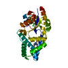
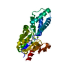
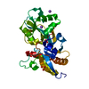
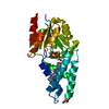
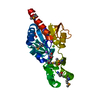
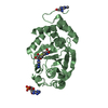
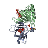
| Structure viewer | Molecule: MolmilJmol/JSmol |
|---|
- Downloads & links
-Download
| PDBx/mmCIF format | 6z1l.cif.gz | 128.1 KB | Display | PDBx/mmCIF format |
|---|---|---|---|---|
| PDB format | pdb6z1l.ent.gz | 84.4 KB | Display | PDB format |
| PDBx/mmJSON format | 6z1l.json.gz | Tree view | PDBx/mmJSON format | |
| Others |  Other downloads Other downloads |
-Validation report
| Arichive directory | https://data.pdbj.org/pub/pdb/validation_reports/z1/6z1lftp://data.pdbj.org/pub/pdb/validation_reports/z1/6z1l | HTTPS FTP |
|---|
-Related structure data
| Related structure data |  6z1kC 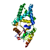 7o1dC 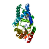 6q7oS S: Starting model for refinement C: citing same article ( |
|---|---|
| Similar structure data |


-Links
 PDBj
PDBj- Assembly
| Deposited unit | 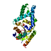
| ||||||||||||
|---|---|---|---|---|---|---|---|---|---|---|---|---|---|
| 1 |
| ||||||||||||
| Unit cell |
|
-Components
| #1: Protein | Mass: 27559.633 Da / Num. of mol.: 1 Source method: isolated from a genetically manipulated source Source: (gene. exp.) synthetic construct (others) / Production host:  Escherichia coli (E. coli) Escherichia coli (E. coli) |
|---|---|
| #2: Chemical | ChemComp-PO4 / Phosphate  Mass: 94.971 Da / Num. of mol.: 1 / Source method: obtained synthetically / Formula: PO4 Mass: 94.971 Da / Num. of mol.: 1 / Source method: obtained synthetically / Formula: PO4 |
| #3: Chemical | ChemComp-EDO / Ethylene glycol  Mass: 62.068 Da / Num. of mol.: 1 / Source method: obtained synthetically / Formula: C2H6O2 Mass: 62.068 Da / Num. of mol.: 1 / Source method: obtained synthetically / Formula: C2H6O2 |
| #4: Water | ChemComp-HOH / Water Mass: 18.015 Da / Num. of mol.: 43 / Source method: isolated from a natural source / Formula: H2O Mass: 18.015 Da / Num. of mol.: 43 / Source method: isolated from a natural source / Formula: H2O |
| Has ligand of interest | N |
-Experimental details
-Experiment
| Experiment | Method: X-RAY DIFFRACTION / Number of used crystals: 1 |
|---|
- Sample preparation
| Crystal | Density Matthews: 3.13 Å3/Da / Density % sol: 60.74 % |
|---|---|
| Crystal grow | Temperature: 277 K / Method: vapor diffusion, sitting drop / pH: 7 / Details: 0.1 M SPG 9.0 25 % w/v PEG 1500 / Temp details: Cold room |
-Data collection
| Diffraction | Mean temperature: 100 K / Serial crystal experiment: N |
|---|---|
| Diffraction source | Source: SYNCHROTRON / Site: Diamond / Beamline: I04 / Wavelength: 0.98 Å |
| Detector | Type: DECTRIS PILATUS3 6M / Detector: PIXEL / Date: Aug 4, 2019 |
| Radiation | Protocol: SINGLE WAVELENGTH / Monochromatic (M) / Laue (L): M / Scattering type: x-ray |
| Radiation wavelength | Wavelength: 0.98 Å / Relative weight: 1 |
| Reflection | Resolution: 2.29→42.71 Å / Num. obs: 16121 / % possible obs: 99.55 % / Redundancy: 9.8 % / Biso Wilson estimate: 65.33 Å2 / CC1/2: 0.999 / CC star: 1 / Rmerge(I) obs: 0.0806 / Rpim(I) all: 0.02701 / Rrim(I) all: 0.08509 / Net I/σ(I): 10.51 |
| Reflection shell | Resolution: 2.29→2.372 Å / Redundancy: 9.6 % / Rmerge(I) obs: 1.617 / Mean I/σ(I) obs: 1.21 / Num. unique obs: 1571 / CC1/2: 0.603 / CC star: 0.867 / Rpim(I) all: 0.5474 / % possible all: 98.66 |
- Processing
| Software |
| |||||||||||||||||||||||||||||||||||||||||||||||||
|---|---|---|---|---|---|---|---|---|---|---|---|---|---|---|---|---|---|---|---|---|---|---|---|---|---|---|---|---|---|---|---|---|---|---|---|---|---|---|---|---|---|---|---|---|---|---|---|---|---|---|
| Refinement | Method to determine structure: MOLECULAR REPLACEMENT Starting model: 6Q7O Resolution: 2.29→42.71 Å / SU ML: 0.3306 / Cross valid method: FREE R-VALUE / σ(F): 1.33 / Phase error: 33.9881 Stereochemistry target values: GeoStd + Monomer Library + CDL v1.2
| |||||||||||||||||||||||||||||||||||||||||||||||||
| Solvent computation | Shrinkage radii: 0.9 Å / VDW probe radii: 1.11 Å / Solvent model: FLAT BULK SOLVENT MODEL | |||||||||||||||||||||||||||||||||||||||||||||||||
| Displacement parameters | Biso mean: 75.73 Å2 | |||||||||||||||||||||||||||||||||||||||||||||||||
| Refinement step | Cycle: LAST / Resolution: 2.29→42.71 Å
| |||||||||||||||||||||||||||||||||||||||||||||||||
| Refine LS restraints |
| |||||||||||||||||||||||||||||||||||||||||||||||||
| LS refinement shell |
| |||||||||||||||||||||||||||||||||||||||||||||||||
| Refinement TLS params. | Method: refined / Origin x: -20.9491046743 Å / Origin y: 26.477902989 Å / Origin z: -11.0134784038 Å
| |||||||||||||||||||||||||||||||||||||||||||||||||
| Refinement TLS group | Selection details: all |
