 Movie
Movie Controller


+ Open data

- Basic information
| Entry | Database: PDB / ID: 6z05 | ||||||
|---|---|---|---|---|---|---|---|
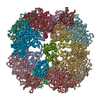
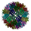
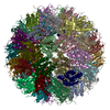
| Title | Campylobacter jejuni serine protease HtrA | ||||||
 Components Components | DegQ family serine endoprotease | ||||||
 Keywords Keywords |  LYASE / Campylobacter jejuni / serine protease / HtrA / dodecamer LYASE / Campylobacter jejuni / serine protease / HtrA / dodecamer | ||||||
| Function / homology |  Function and homology information Function and homology information | ||||||
| Biological species |   Campylobacter jejuni (Campylobacter) Campylobacter jejuni (Campylobacter) | ||||||
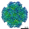
| Method | ELECTRON MICROSCOPY / single particle reconstruction / cryo EM / Resolution: 5.8 Å | ||||||
 Authors Authors | Grinzato, A. / Kandiah, E. / Zanotti, G. | ||||||
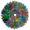
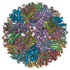
 Citation Citation | Journal: Gut Microbes / Year: 2020 Title: Functional analysis and cryo-electron microscopy of serine protease HtrA. Authors: Urszula Zarzecka / Alessandro Grinzato / Eaazhisai Kandiah / Dominik Cysewski / Paola Berto / Joanna Skorko-Glonek / Giuseppe Zanotti / Steffen Backert /     Abstract: is a predominant zoonotic pathogen causing gastroenteritis and other diseases in humans. An important bacterial virulence factor is the secreted serine protease HtrA (HtrA ), which targets tight ... is a predominant zoonotic pathogen causing gastroenteritis and other diseases in humans. An important bacterial virulence factor is the secreted serine protease HtrA (HtrA ), which targets tight and adherens junctional proteins in the gut epithelium. Here we have investigated the function and structure of HtrA using biochemical assays and cryo-electron microscopy. Mass spectrometry analysis identified differences and similarities in the cleavage site specificity for HtrA by comparison to the HtrA counterparts from and . We defined the architecture of HtrA at 5.8 Å resolution as a dodecamer, built of four trimers. The contacts between the trimers are quite loose, a fact that explains the flexibility and mobility of the dodecameric assembly. This flexibility has also been studied through molecular dynamics simulation, which revealed opening of the dodecamer to expose the proteolytically active site of the protease. Moreover, we examined the rearrangements at the level of oligomerization in the presence or absence of substrate using size exclusion chromatography, which revealed hexamers, dodecamers and larger oligomeric forms, as well as remarkable stability of higher oligomeric forms (> 12-mers) compared to previously tested homologs from other bacteria. Extremely dynamic decay of the higher oligomeric forms into lower forms was observed after full cleavage of the substrate by the proteolytically active variant of HtrA . Together, this is the first report on the in-depth functional and structural analysis of HtrA , which may allow the construction of therapeutically relevant HtrA inhibitors in the near future. | ||||||
| History |
|
- Structure visualization
| Movie |
Movie viewer |
|---|---|
| Structure viewer | Molecule: MolmilJmol/JSmol |
- Downloads & links
-Download
| PDBx/mmCIF format | 6z05.cif.gz | 807.8 KB | Display | PDBx/mmCIF format |
|---|---|---|---|---|
| PDB format | pdb6z05.ent.gz | 674.5 KB | Display | PDB format |
| PDBx/mmJSON format | 6z05.json.gz | Tree view | PDBx/mmJSON format | |
| Others |  Other downloads Other downloads |
-Validation report
| Arichive directory | https://data.pdbj.org/pub/pdb/validation_reports/z0/6z05ftp://data.pdbj.org/pub/pdb/validation_reports/z0/6z05 | HTTPS FTP |
|---|
-Related structure data
| Related structure data | 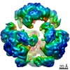 11003MC M: map data used to model this data C: citing same article ( |
|---|---|
| Similar structure data |
-Links
 PDBj
PDBj
- Assembly
| Deposited unit | 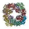
|
|---|---|
| 1 |
|
-Components
| #1: Protein | Mass: 47182.379 Da / Num. of mol.: 12 Source method: isolated from a genetically manipulated source Source: (gene. exp.) Campylobacter jejuni (Campylobacter)Gene: htrA, AY595_01940, B7T14_05270, BB943_00930, BB980_02965, BOI95_01210, CA962_01090, CKH46_05385, CW563_00090, DK813_04915, DQX79_02990, DUH84_04555, DW530_01050, F0N90_05545, F1O60_00260, F3L27_ ...Gene: htrA, AY595_01940, B7T14_05270, BB943_00930, BB980_02965, BOI95_01210, CA962_01090, CKH46_05385, CW563_00090, DK813_04915, DQX79_02990, DUH84_04555, DW530_01050, F0N90_05545, F1O60_00260, F3L27_00375, F6319_03730, F7858_01545, FR416_00510, FRM19_03410, FRQ83_01860, FZ422_01615, FZU65_03575 Production host: Escherichia coli (E. coli) / References: UniProt: A0A620LS18 |
|---|
-Experimental details
-Experiment
| Experiment | Method: ELECTRON MICROSCOPY |
|---|---|
| EM experiment | Aggregation state: PARTICLE / 3D reconstruction method: single particle reconstruction |
- Sample preparation
| Component | Name: HtrA dodecamer / Type: COMPLEX / Entity ID: all / Source: RECOMBINANT |
|---|---|
| Molecular weight | Experimental value: NO |
| Source (natural) | Organism: Campylobacter jejuni (Campylobacter) |
| Source (recombinant) | Organism: Escherichia coli (E. coli) |
| Buffer solution | pH: 7.4 |
| Specimen | Embedding applied: NO / Shadowing applied: NO / Staining applied: NO / Vitrification applied: YES |
| Vitrification | Cryogen name: ETHANE |
- Electron microscopy imaging
| Microscopy | Model: TFS GLACIOS |
|---|---|
| Electron gun | Electron source: FIELD EMISSION GUN / Accelerating voltage: 200 kV / Illumination mode: FLOOD BEAM |
| Electron lens | Mode: BRIGHT FIELDBright-field microscopy |
| Image recording | Electron dose: 44 e/Å2 / Film or detector model: FEI FALCON III (4k x 4k) |
- Processing
| CTF correction | Type: NONE |
|---|---|
| Symmetry | Point symmetry: T (tetrahedral) |
| 3D reconstruction | Resolution: 5.8 Å / Resolution method: FSC 0.143 CUT-OFF / Num. of particles: 113843 / Symmetry type: POINT |
