 Movie
Movie Controller

[English] 日本語
 Yorodumi
- PDB-6yqb: Taka-amylase in complex with alpha-glucosyl epi-cyclophellitol cy... -
+ Open data

- Basic information
| Entry | Database: PDB / ID: 6yqb | ||||||
|---|---|---|---|---|---|---|---|
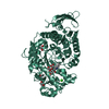
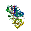
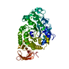
| Title | Taka-amylase in complex with alpha-glucosyl epi-cyclophellitol cyclosulfate inhibitor | ||||||
 Components Components | Alpha-amylase | ||||||
 Keywords Keywords | HYDROLASE / Inhibitor / Complex / Amylase | ||||||
| Function / homology |  Function and homology informationalpha-amylase / carbohydrate catabolic process / alpha-amylase activity / calcium ion binding Function and homology informationalpha-amylase / carbohydrate catabolic process / alpha-amylase activity / calcium ion bindingSimilarity search - Function | ||||||
| Biological species |  Aspergillus oryzae (mold) Aspergillus oryzae (mold) | ||||||
| Method | X-RAY DIFFRACTION / SYNCHROTRON / MOLECULAR REPLACEMENT / Resolution: 1.5 Å | ||||||
 Authors Authors | Armstrong, Z. / Chen, Y. / Artola, M. / Overkleeft, H. / Davies, G. | ||||||
| Funding support |  United Kingdom, 1items United Kingdom, 1items
| ||||||
 Citation Citation | Journal: J.Am.Chem.Soc. / Year: 2021 Title: Activity-Based Protein Profiling of Retaining alpha-Amylases in Complex Biological Samples. Authors: Chen, Y. / Armstrong, Z. / Artola, M. / Florea, B.I. / Kuo, C.L. / de Boer, C. / Rasmussen, M.S. / Abou Hachem, M. / van der Marel, G.A. / Codee, J.D.C. / Aerts, J.M.F.G. / Davies, G.J. / Overkleeft, H.S. | ||||||
| History |
|
- Structure visualization
| Structure viewer | Molecule: MolmilJmol/JSmol |
|---|
- Downloads & links
-Download
| PDBx/mmCIF format | 6yqb.cif.gz | 364.6 KB | Display | PDBx/mmCIF format |
|---|---|---|---|---|
| PDB format | pdb6yqb.ent.gz | Display | PDB format | |
| PDBx/mmJSON format | 6yqb.json.gz | Tree view | PDBx/mmJSON format | |
| Others |  Other downloads Other downloads |
-Validation report
| Arichive directory | https://data.pdbj.org/pub/pdb/validation_reports/yq/6yqbftp://data.pdbj.org/pub/pdb/validation_reports/yq/6yqb | HTTPS FTP |
|---|
-Related structure data
| Related structure data |  6yq7C  6yq9C  6yqaC  6yqcC 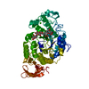 7taaS S: Starting model for refinement C: citing same article ( |
|---|---|
| Similar structure data |


-Links
 PDBj
PDBj


- Assembly
| Deposited unit | 
| ||||||||
|---|---|---|---|---|---|---|---|---|---|
| 1 | 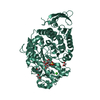
| ||||||||
| 2 | 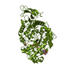
| ||||||||
| Unit cell |
|
-Components
-Protein , 1 types, 2 molecules AAABBB
| #1: Protein | Mass: 54846.762 Da / Num. of mol.: 2 Source method: isolated from a genetically manipulated source Source: (gene. exp.) Aspergillus oryzae (mold) / Gene: OAory_01097160 / Production host: Aspergillus oryzae (mold) / References: UniProt: A0A1S9DH83, alpha-amylase |
|---|
-Sugars , 2 types, 4 molecules 

| #3: Sugar | N-Acetylglucosamine Type: D-saccharide, beta linking / Mass: 221.208 Da / Num. of mol.: 2 Type: D-saccharide, beta linking / Mass: 221.208 Da / Num. of mol.: 2Source method: isolated from a genetically manipulated source Formula: C8H15NO6 #5: Sugar | Glucose Type: D-saccharide, alpha linking / Mass: 180.156 Da / Num. of mol.: 2 Type: D-saccharide, alpha linking / Mass: 180.156 Da / Num. of mol.: 2Source method: isolated from a genetically manipulated source Formula: C6H12O6 |
|---|
-Non-polymers , 4 types, 795 molecules 


| #2: Chemical | ChemComp-EDO / Ethylene glycol Mass: 62.068 Da / Num. of mol.: 6 / Source method: obtained synthetically / Formula: C2H6O2 Mass: 62.068 Da / Num. of mol.: 6 / Source method: obtained synthetically / Formula: C2H6O2#4: Chemical |  Mass: 40.078 Da / Num. of mol.: 2 / Source method: obtained synthetically / Formula: Ca Mass: 40.078 Da / Num. of mol.: 2 / Source method: obtained synthetically / Formula: Ca#6: Chemical |  Mass: 274.246 Da / Num. of mol.: 2 / Source method: obtained synthetically / Formula: C7H14O9S Mass: 274.246 Da / Num. of mol.: 2 / Source method: obtained synthetically / Formula: C7H14O9S#7: Water | ChemComp-HOH / | WaterMass: 18.015 Da / Num. of mol.: 785 / Source method: isolated from a natural source / Formula: H2O |
|---|
-Details
| Has ligand of interest | Y |
|---|
-Experimental details
-Experiment
| Experiment | Method: X-RAY DIFFRACTION / Number of used crystals: 1 |
|---|
- Sample preparation
| Crystal | Density Matthews: 2.26 Å3/Da / Density % sol: 45.57 % |
|---|---|
| Crystal grow | Temperature: 293 K / Method: vapor diffusion, sitting drop / Details: 0.1 M sodium citrate pH 5.6, 18% (v/v) 2-propanol |
-Data collection
| Diffraction | Mean temperature: 100 K / Serial crystal experiment: N |
|---|---|
| Diffraction source | Source: SYNCHROTRON / Site: Diamond / Beamline: I04 / Wavelength: 0.979499 Å |
| Detector | Type: DECTRIS EIGER2 X 16M / Detector: PIXEL / Date: Mar 7, 2019 |
| Radiation | Protocol: SINGLE WAVELENGTH / Monochromatic (M) / Laue (L): M / Scattering type: x-ray |
| Radiation wavelength | Wavelength: 0.979499 Å / Relative weight: 1 |
| Reflection | Resolution: 1.5→59.78 Å / Num. obs: 153290 / % possible obs: 98.7 % / Redundancy: 6.8 % / CC1/2: 0.999 / Net I/σ(I): 10.3 |
| Reflection shell | Resolution: 1.5→1.53 Å / Num. unique obs: 7423 / CC1/2: 0.917 |
- Processing
| Software |
| ||||||||||||||||||||||||||||||||||||||||||||||||||||||||||||||||||||||||||||||||||||||||||||||||||||||||||||||||||||||||||||||||||||||||||||||||||||||||||||||||
|---|---|---|---|---|---|---|---|---|---|---|---|---|---|---|---|---|---|---|---|---|---|---|---|---|---|---|---|---|---|---|---|---|---|---|---|---|---|---|---|---|---|---|---|---|---|---|---|---|---|---|---|---|---|---|---|---|---|---|---|---|---|---|---|---|---|---|---|---|---|---|---|---|---|---|---|---|---|---|---|---|---|---|---|---|---|---|---|---|---|---|---|---|---|---|---|---|---|---|---|---|---|---|---|---|---|---|---|---|---|---|---|---|---|---|---|---|---|---|---|---|---|---|---|---|---|---|---|---|---|---|---|---|---|---|---|---|---|---|---|---|---|---|---|---|---|---|---|---|---|---|---|---|---|---|---|---|---|---|---|---|---|
| Refinement | Method to determine structure: MOLECULAR REPLACEMENT Starting model: 7taa Resolution: 1.5→59.78 Å / Cor.coef. Fo:Fc: 0.978 / Cor.coef. Fo:Fc free: 0.969 / Cross valid method: FREE R-VALUE / ESU R: 0.066 / ESU R Free: 0.068 Details: Hydrogens have been added in their riding positions
| ||||||||||||||||||||||||||||||||||||||||||||||||||||||||||||||||||||||||||||||||||||||||||||||||||||||||||||||||||||||||||||||||||||||||||||||||||||||||||||||||
| Solvent computation | Ion probe radii: 0.8 Å / Shrinkage radii: 0.8 Å / VDW probe radii: 1.2 Å | ||||||||||||||||||||||||||||||||||||||||||||||||||||||||||||||||||||||||||||||||||||||||||||||||||||||||||||||||||||||||||||||||||||||||||||||||||||||||||||||||
| Displacement parameters | Biso mean: 20.58 Å2
| ||||||||||||||||||||||||||||||||||||||||||||||||||||||||||||||||||||||||||||||||||||||||||||||||||||||||||||||||||||||||||||||||||||||||||||||||||||||||||||||||
| Refinement step | Cycle: LAST / Resolution: 1.5→59.78 Å
| ||||||||||||||||||||||||||||||||||||||||||||||||||||||||||||||||||||||||||||||||||||||||||||||||||||||||||||||||||||||||||||||||||||||||||||||||||||||||||||||||
| Refine LS restraints |
| ||||||||||||||||||||||||||||||||||||||||||||||||||||||||||||||||||||||||||||||||||||||||||||||||||||||||||||||||||||||||||||||||||||||||||||||||||||||||||||||||
| LS refinement shell |
|
