 Movie
Movie Controller


+ Open data

- Basic information
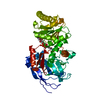
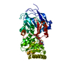
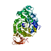
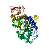
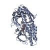
| Entry | Database: PDB / ID: 1oyh | |||||||||
|---|---|---|---|---|---|---|---|---|---|---|
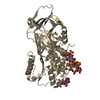
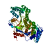
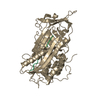
| Title | Crystal Structure of P13 Alanine Variant of Antithrombin | |||||||||
 Components Components | (Antithrombin- ...) x 2 | |||||||||
 Keywords Keywords |  BLOOD CLOTTING / thrombin / inhibition / heparin analogue / SERINE PROTEASE INHIBITOR BLOOD CLOTTING / thrombin / inhibition / heparin analogue / SERINE PROTEASE INHIBITOR | |||||||||
| Function / homology |  Function and homology informationregulation of blood coagulation / Common Pathway of Fibrin Clot Formation / Intrinsic Pathway of Fibrin Clot Formation / Post-translational protein phosphorylation / serine-type endopeptidase inhibitor activity / Regulation of Insulin-like Growth Factor (IGF) transport and uptake by Insulin-like Growth Factor Binding Proteins (IGFBPs) / blood coagulation / heparin binding / collagen-containing extracellular matrix / protease binding ...regulation of blood coagulation / Common Pathway of Fibrin Clot Formation / Intrinsic Pathway of Fibrin Clot Formation / Post-translational protein phosphorylation / serine-type endopeptidase inhibitor activity / Regulation of Insulin-like Growth Factor (IGF) transport and uptake by Insulin-like Growth Factor Binding Proteins (IGFBPs) / blood coagulation / heparin binding / collagen-containing extracellular matrix / protease binding / blood microparticle / endoplasmic reticulum lumen / extracellular space / extracellular exosome / extracellular region / identical protein binding / plasma membrane Function and homology informationregulation of blood coagulation / Common Pathway of Fibrin Clot Formation / Intrinsic Pathway of Fibrin Clot Formation / Post-translational protein phosphorylation / serine-type endopeptidase inhibitor activity / Regulation of Insulin-like Growth Factor (IGF) transport and uptake by Insulin-like Growth Factor Binding Proteins (IGFBPs) / blood coagulation / heparin binding / collagen-containing extracellular matrix / protease binding ...regulation of blood coagulation / Common Pathway of Fibrin Clot Formation / Intrinsic Pathway of Fibrin Clot Formation / Post-translational protein phosphorylation / serine-type endopeptidase inhibitor activity / Regulation of Insulin-like Growth Factor (IGF) transport and uptake by Insulin-like Growth Factor Binding Proteins (IGFBPs) / blood coagulation / heparin binding / collagen-containing extracellular matrix / protease binding / blood microparticle / endoplasmic reticulum lumen / extracellular space / extracellular exosome / extracellular region / identical protein binding / plasma membraneSimilarity search - Function | |||||||||
| Biological species |  Homo sapiens (human) Homo sapiens (human) | |||||||||
| Method | X-RAY DIFFRACTION / MOLECULAR REPLACEMENT / Resolution: 2.62 Å | |||||||||
 Authors Authors | Johnson, D.J.D. / Huntington, J.A. | |||||||||
 Citation Citation | Journal: J.Biol.Chem. / Year: 2004 Title: The influence of hinge region residue Glu-381 on antithrombin allostery and metastability Authors: Johnson, D.J.D. / Huntington, J.A. | |||||||||
| History |
|
- Structure visualization
| Structure viewer | Molecule: MolmilJmol/JSmol |
|---|
- Downloads & links
-Download
| PDBx/mmCIF format | 1oyh.cif.gz | 179.1 KB | Display | PDBx/mmCIF format |
|---|---|---|---|---|
| PDB format | pdb1oyh.ent.gz | 140.2 KB | Display | PDB format |
| PDBx/mmJSON format | 1oyh.json.gz | Tree view | PDBx/mmJSON format | |
| Others |  Other downloads Other downloads |
-Validation report
| Arichive directory | https://data.pdbj.org/pub/pdb/validation_reports/oy/1oyhftp://data.pdbj.org/pub/pdb/validation_reports/oy/1oyh | HTTPS FTP |
|---|
-Related structure data
| Related structure data | 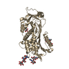 1e04S S: Starting model for refinement |
|---|---|
| Similar structure data |


-Links
 PDBj
PDBj


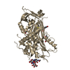
- Assembly
| Deposited unit | 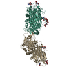
| ||||||||||
|---|---|---|---|---|---|---|---|---|---|---|---|
| 1 | 
| ||||||||||
| 2 | 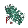
| ||||||||||
| Unit cell |
|
-Components
-Antithrombin- ... , 2 types, 2 molecules IL
| #1: Protein | / ATIII / PRO0309 Mass: 49026.980 Da / Num. of mol.: 1 / Mutation: E381A, S137A Source method: isolated from a genetically manipulated source Details: E381A mutant on the beta glycoform background of S137A Source: (gene. exp.) Homo sapiens (human) / Tissue: plasmaDescription: Plasma alpha antithrombin was converted to latent by the glycerol method, and is chain L Organ: blood / Plasmid: pMASTOP / Cell (production host): Baby Hampster Kidney Cells / Production host:   Cricetulus griseus (Chinese hamster) / References: UniProt: P01008 Cricetulus griseus (Chinese hamster) / References: UniProt: P01008 |
|---|---|
| #2: Protein | / ATIII / PRO0309 Mass: 49101.016 Da / Num. of mol.: 1 Source method: isolated from a genetically manipulated source Details: beta glycoform / Source: (gene. exp.) Homo sapiens (human) / Tissue: plasmaDescription: Plasma alpha antithrombin was converted to latent by the glycerol method, and is chain L Organ: blood / Plasmid: pMASTOP / Cell (production host): Baby Hampster Kidney Cells / Production host: Cricetulus griseus (Chinese hamster) / References: UniProt: P01008 |
-Sugars , 3 types, 7 molecules 
| #3: Polysaccharide | / Mass: 424.401 Da / Num. of mol.: 2 Source method: isolated from a genetically manipulated source #4: Polysaccharide | alpha-D-mannopyranose-(1-3)-[alpha-D-mannopyranose-(1-6)]beta-D-mannopyranose-(1-4)-2-acetamido-2- ...alpha-D-mannopyranose-(1-3)-[alpha-D-mannopyranose-(1-6)]beta-D-mannopyranose-(1-4)-2-acetamido-2-deoxy-beta-D-glucopyranose-(1-4)-2-acetamido-2-deoxy-beta-D-glucopyranose | / Mass: 910.823 Da / Num. of mol.: 1Source method: isolated from a genetically manipulated source #5: Sugar | ChemComp-NAG / N-Acetylglucosamine Type: D-saccharide, beta linking / Mass: 221.208 Da / Num. of mol.: 4 Type: D-saccharide, beta linking / Mass: 221.208 Da / Num. of mol.: 4Source method: isolated from a genetically manipulated source Formula: C8H15NO6 |
|---|
-Non-polymers , 1 types, 54 molecules 
| #6: Water | ChemComp-HOH / WaterMass: 18.015 Da / Num. of mol.: 54 / Source method: isolated from a natural source / Formula: H2O |
|---|
-Experimental details
-Experiment
| Experiment | Method: X-RAY DIFFRACTION / Number of used crystals: 1 |
|---|
- Sample preparation
| Crystal | Density Matthews: 3.14 Å3/Da / Density % sol: 60.5 % |
|---|---|
| Crystal grow | Temperature: 293 K / Method: vapor diffusion, hanging drop / pH: 6.7 Details: Na/K Phosphate, PEG 4000, pH 6.7, VAPOR DIFFUSION, HANGING DROP, temperature 293K |
-Data collection
| Diffraction | Mean temperature: 100 K |
|---|---|
| Diffraction source | Source: ROTATING ANODE / Type: RIGAKU / Wavelength: 1.5418 Å |
| Detector | Detector: AREA DETECTOR |
| Radiation | Protocol: SINGLE WAVELENGTH / Monochromatic (M) / Laue (L): M / Scattering type: x-ray |
| Radiation wavelength | Wavelength: 1.5418 Å / Relative weight: 1 |
| Reflection | Resolution: 2.62→39.5 Å / Num. all: 36632 / Num. obs: 35267 / % possible obs: 99.4 % / Observed criterion σ(I): -3 / Redundancy: 6.1 % / Biso Wilson estimate: 76.609 Å2 / Rmerge(I) obs: 0.073 / Rsym value: 0.067 / Net I/σ(I): 9 |
| Reflection shell | Resolution: 2.62→2.74 Å / Redundancy: 5.9 % / Rmerge(I) obs: 0.496 / Mean I/σ(I) obs: 3.2 / Num. unique all: 5109 / Rsym value: 0.453 / % possible all: 96.4 |
- Processing
| Software |
| |||||||||||||||||||||||||
|---|---|---|---|---|---|---|---|---|---|---|---|---|---|---|---|---|---|---|---|---|---|---|---|---|---|---|
| Refinement | Method to determine structure: MOLECULAR REPLACEMENT / Starting model: 10000 / Resolution: 2.62→39 Å / Isotropic thermal model: restrained / Cross valid method: THROUGHOUT / σ(F): 0 / Stereochemistry target values: Engh & Huber / Details: used mlf target
| |||||||||||||||||||||||||
| Displacement parameters |
| |||||||||||||||||||||||||
| Refine analyze |
| |||||||||||||||||||||||||
| Refinement step | Cycle: LAST / Resolution: 2.62→39 Å
| |||||||||||||||||||||||||
| Refine LS restraints |
| |||||||||||||||||||||||||
| LS refinement shell | Resolution: 2.62→2.76 Å / Rfactor Rfree error: 0.023
|
