 Movie
Movie Controller


+ Open data

- Basic information
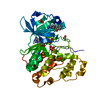
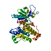
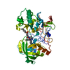
| Entry | Database: PDB / ID: 6ypk | ||||||
|---|---|---|---|---|---|---|---|
| Title | Crystal Structure of CK2alpha with GTP bound | ||||||
 Components Components | Casein kinase II subunit alpha Casein kinase 2 Casein kinase 2 | ||||||
 Keywords Keywords | TRANSFERASE / selective ATP competitive inhibitors | ||||||
| Function / homology |  Function and homology information Function and homology informationregulation of chromosome separation / positive regulation of aggrephagy / WNT mediated activation of DVL / Condensation of Prometaphase Chromosomes / protein kinase CK2 complex / symbiont-mediated disruption of host cell PML body / Receptor Mediated Mitophagy / Sin3-type complex / Synthesis of PC / RUNX1 interacts with co-factors whose precise effect on RUNX1 targets is not known ...regulation of chromosome separation / positive regulation of aggrephagy / WNT mediated activation of DVL / Condensation of Prometaphase Chromosomes / protein kinase CK2 complex / symbiont-mediated disruption of host cell PML body / Receptor Mediated Mitophagy / Sin3-type complex / Synthesis of PC / RUNX1 interacts with co-factors whose precise effect on RUNX1 targets is not known / negative regulation of apoptotic signaling pathway / positive regulation of Wnt signaling pathway / chaperone-mediated protein folding / negative regulation of ubiquitin-dependent protein catabolic process / Signal transduction by L1 / peptidyl-threonine phosphorylation / Hsp90 protein binding / negative regulation of cysteine-type endopeptidase activity involved in apoptotic process / PML body / Wnt signaling pathway / Regulation of PTEN stability and activity / Cooperation of PDCL (PhLP1) and TRiC/CCT in G-protein beta folding / positive regulation of protein catabolic process / rhythmic process / KEAP1-NFE2L2 pathway / double-strand break repair / kinase activity / positive regulation of cell growth / peptidyl-serine phosphorylation / Regulation of TP53 Activity through Phosphorylation / negative regulation of translation / protein stabilization / regulation of cell cycle / non-specific serine/threonine protein kinase / cell cycle / protein phosphorylation / protein serine kinase activity / protein serine/threonine kinase activity / apoptotic process / DNA damage response / positive regulation of cell population proliferation / signal transduction / nucleoplasm / ATP binding / identical protein binding / nucleus / plasma membrane / cytosolSimilarity search - Function | ||||||
| Biological species |  Homo sapiens (human) Homo sapiens (human) | ||||||
| Method | X-RAY DIFFRACTION / SYNCHROTRON / MOLECULAR REPLACEMENT / molecular replacement / Resolution: 1.79 Å | ||||||
 Authors Authors | Brear, P. / Hyvonen, M. | ||||||
 Citation Citation | Journal: J.Med.Chem. / Year: 2020 Title: Proposed Allosteric Inhibitors Bind to the ATP Site of CK2 alpha. Authors: Brear, P. / Ball, D. / Stott, K. / D'Arcy, S. / Hyvonen, M. | ||||||
| History |
|
- Structure visualization
| Structure viewer | Molecule: MolmilJmol/JSmol |
|---|
- Downloads & links
-Download
| PDBx/mmCIF format | 6ypk.cif.gz | 88.6 KB | Display | PDBx/mmCIF format |
|---|---|---|---|---|
| PDB format | pdb6ypk.ent.gz | 64.5 KB | Display | PDB format |
| PDBx/mmJSON format | 6ypk.json.gz | Tree view | PDBx/mmJSON format | |
| Others |  Other downloads Other downloads |
-Validation report
| Arichive directory | https://data.pdbj.org/pub/pdb/validation_reports/yp/6ypkftp://data.pdbj.org/pub/pdb/validation_reports/yp/6ypk | HTTPS FTP |
|---|
-Related structure data
| Related structure data |  6ypgC  6yphC  6ypjC  6ypnC  5cvhS S: Starting model for refinement C: citing same article ( |
|---|---|
| Similar structure data |

-Links
 PDBj
PDBj


- Assembly
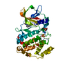
| Deposited unit | 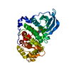
| ||||||||
|---|---|---|---|---|---|---|---|---|---|
| 1 |
| ||||||||
| Unit cell |
|
-Components
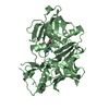
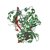
| #1: Protein | Casein kinase 2 / CK II alpha Mass: 39031.391 Da / Num. of mol.: 1 / Mutation: K75A Source method: isolated from a genetically manipulated source Source: (gene. exp.) Homo sapiens (human) / Gene: CSNK2A1, CK2A1 / Plasmid: pHAT2 / Production host:  Escherichia coli BL21(DE3) (bacteria) Escherichia coli BL21(DE3) (bacteria)References: UniProt: P68400, non-specific serine/threonine protein kinase |
|---|---|
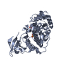
| #2: Chemical | ChemComp-GDP / Guanosine diphosphate  Type: RNA linking / Mass: 443.201 Da / Num. of mol.: 1 / Source method: obtained synthetically / Formula: C10H15N5O11P2 / Feature type: SUBJECT OF INVESTIGATION / Comment: GDP, energy-carrying molecule*YM Type: RNA linking / Mass: 443.201 Da / Num. of mol.: 1 / Source method: obtained synthetically / Formula: C10H15N5O11P2 / Feature type: SUBJECT OF INVESTIGATION / Comment: GDP, energy-carrying molecule*YM |
| #3: Water | ChemComp-HOH / Water Mass: 18.015 Da / Num. of mol.: 144 / Source method: isolated from a natural source / Formula: H2O Mass: 18.015 Da / Num. of mol.: 144 / Source method: isolated from a natural source / Formula: H2O |
| Has ligand of interest | Y |
-Experimental details
-Experiment
| Experiment | Method: X-RAY DIFFRACTION / Number of used crystals: 1 |
|---|
- Sample preparation
| Crystal | Density Matthews: 1.99 Å3/Da / Density % sol: 38.13 % / Mosaicity: 0 ° |
|---|---|
| Crystal grow | Temperature: 298 K / Method: vapor diffusion, sitting drop / pH: 6.5 Details: 112.5mM Mes, 35% glycerol ethoxylate, 180 mM ammonium acetate |
-Data collection
| Diffraction | Mean temperature: 100 K / Serial crystal experiment: N | ||||||||||||||||||||||||||||||
|---|---|---|---|---|---|---|---|---|---|---|---|---|---|---|---|---|---|---|---|---|---|---|---|---|---|---|---|---|---|---|---|
| Diffraction source | Source: SYNCHROTRON / Site: Diamond  / Beamline: I24 / Wavelength: 0.9686 Å / Beamline: I24 / Wavelength: 0.9686 Å | ||||||||||||||||||||||||||||||
| Detector | Type: DECTRIS PILATUS3 6M / Detector: PIXEL / Date: Sep 25, 2016 | ||||||||||||||||||||||||||||||
| Radiation | Protocol: SINGLE WAVELENGTH / Monochromatic (M) / Laue (L): M / Scattering type: x-ray | ||||||||||||||||||||||||||||||
| Radiation wavelength | Wavelength: 0.9686 Å / Relative weight: 1 | ||||||||||||||||||||||||||||||
| Reflection | Resolution: 1.79→49.7 Å / Num. obs: 28931 / % possible obs: 99.2 % / Redundancy: 3.3 % / Biso Wilson estimate: 29.95 Å2 / CC1/2: 0.982 / Rmerge(I) obs: 0.138 / Rpim(I) all: 0.09 / Rrim(I) all: 0.165 / Net I/σ(I): 6.5 / Num. measured all: 94177 | ||||||||||||||||||||||||||||||
| Reflection shell | Diffraction-ID: 1
|
-Phasing
| Phasing | Method: molecular replacement |
|---|
- Processing
| Software |
| ||||||||||||||||||||||||||||||||||||||||||||||||||||||||||||||||||||||||||||||||||||||||||||||||||||||||||||
|---|---|---|---|---|---|---|---|---|---|---|---|---|---|---|---|---|---|---|---|---|---|---|---|---|---|---|---|---|---|---|---|---|---|---|---|---|---|---|---|---|---|---|---|---|---|---|---|---|---|---|---|---|---|---|---|---|---|---|---|---|---|---|---|---|---|---|---|---|---|---|---|---|---|---|---|---|---|---|---|---|---|---|---|---|---|---|---|---|---|---|---|---|---|---|---|---|---|---|---|---|---|---|---|---|---|---|---|---|---|
| Refinement | Method to determine structure: MOLECULAR REPLACEMENT Starting model: 5CVH Resolution: 1.79→22.31 Å / Cor.coef. Fo:Fc: 0.948 / Cor.coef. Fo:Fc free: 0.934 / SU R Cruickshank DPI: 0.134 / Cross valid method: THROUGHOUT / σ(F): 0 / SU R Blow DPI: 0.133 / SU Rfree Blow DPI: 0.119 / SU Rfree Cruickshank DPI: 0.12
| ||||||||||||||||||||||||||||||||||||||||||||||||||||||||||||||||||||||||||||||||||||||||||||||||||||||||||||
| Displacement parameters | Biso max: 119.58 Å2 / Biso mean: 34.21 Å2 / Biso min: 15.11 Å2
| ||||||||||||||||||||||||||||||||||||||||||||||||||||||||||||||||||||||||||||||||||||||||||||||||||||||||||||
| Refinement step | Cycle: final / Resolution: 1.79→22.31 Å
| ||||||||||||||||||||||||||||||||||||||||||||||||||||||||||||||||||||||||||||||||||||||||||||||||||||||||||||
| Refine LS restraints |
| ||||||||||||||||||||||||||||||||||||||||||||||||||||||||||||||||||||||||||||||||||||||||||||||||||||||||||||
| LS refinement shell | Resolution: 1.79→1.85 Å / Rfactor Rfree error: 0 / Total num. of bins used: 15
|
