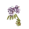
 Movie
Movie Controller


+ Open data

- Basic information
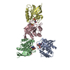
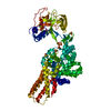
| Entry | Database: PDB / ID: 6yk8 | ||||||
|---|---|---|---|---|---|---|---|
| Title | OTU-like deubiquitinase from Legionella -Lpg2529 | ||||||
 Components Components | Uncharacterized protein | ||||||
 Keywords Keywords | CELL INVASION /  deubiquitinase / legionella / OTU / effector protein deubiquitinase / legionella / OTU / effector protein | ||||||
| Function / homology | : / Dot/Icm T4SS effector Function and homology information Function and homology information | ||||||
| Biological species |  Legionella pneumophila subsp. pneumophila (bacteria) Legionella pneumophila subsp. pneumophila (bacteria) | ||||||
| Method | X-RAY DIFFRACTION / SYNCHROTRON / SAD / Resolution: 2.42 Å | ||||||
 Authors Authors | Shin, D. / Dikic, I. | ||||||
 Citation Citation | Journal: Elife / Year: 2020 Title: Bacterial OTU deubiquitinases regulate substrate ubiquitination upon Legionella infection. Authors: Shin, D. / Bhattacharya, A. / Cheng, Y.L. / Alonso, M.C. / Mehdipour, A.R. / van der Heden van Noort, G.J. / Ovaa, H. / Hummer, G. / Dikic, I. | ||||||
| History |
|
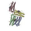
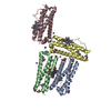
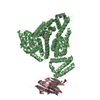
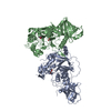
- Structure visualization
| Structure viewer | Molecule: MolmilJmol/JSmol |
|---|
- Downloads & links
-Download
| PDBx/mmCIF format | 6yk8.cif.gz | 143.1 KB | Display | PDBx/mmCIF format |
|---|---|---|---|---|
| PDB format | pdb6yk8.ent.gz | 94.1 KB | Display | PDB format |
| PDBx/mmJSON format | 6yk8.json.gz | Tree view | PDBx/mmJSON format | |
| Others |  Other downloads Other downloads |
-Validation report
| Arichive directory | https://data.pdbj.org/pub/pdb/validation_reports/yk/6yk8ftp://data.pdbj.org/pub/pdb/validation_reports/yk/6yk8 | HTTPS FTP |
|---|
-Related structure data
| Similar structure data |
|---|


-Links
 PDBj
PDBj
- Assembly
| Deposited unit | 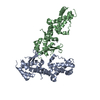
| ||||||||||||
|---|---|---|---|---|---|---|---|---|---|---|---|---|---|
| 1 |
| ||||||||||||
| Unit cell |
|
-Components
| #1: Protein | Mass: 34752.082 Da / Num. of mol.: 2 Source method: isolated from a genetically manipulated source Source: (gene. exp.) Legionella pneumophila subsp. pneumophila (strain Philadelphia 1 / ATCC 33152 / DSM 7513) (bacteria)Strain: Philadelphia 1 / ATCC 33152 / DSM 7513 / Gene: lpg2529 / Production host: Escherichia coli (E. coli) / References: UniProt: Q5ZSI8#2: Chemical | ChemComp-PT /   Mass: 195.078 Da / Num. of mol.: 10 / Source method: obtained synthetically / Formula: Pt Mass: 195.078 Da / Num. of mol.: 10 / Source method: obtained synthetically / Formula: Pt#3: Water | ChemComp-HOH / | Water Mass: 18.015 Da / Num. of mol.: 1 / Source method: isolated from a natural source / Formula: H2O Mass: 18.015 Da / Num. of mol.: 1 / Source method: isolated from a natural source / Formula: H2OHas ligand of interest | N | |
|---|
-Experimental details
-Experiment
| Experiment | Method: X-RAY DIFFRACTION / Number of used crystals: 1 |
|---|
- Sample preparation
| Crystal | Density Matthews: 2.24 Å3/Da / Density % sol: 45.2 % |
|---|---|
| Crystal grow | Temperature: 293 K / Method: vapor diffusion, sitting drop / pH: 8.5 / Details: 25 % PEG3350, 100 mM Tris-HCl pH 8.5, 200 mM NaCl |
-Data collection
| Diffraction | Mean temperature: 100 K / Serial crystal experiment: N |
|---|---|
| Diffraction source | Source: SYNCHROTRON / Site: SLS  / Beamline: X06SA / Wavelength: 1.070333 Å / Beamline: X06SA / Wavelength: 1.070333 Å |
| Detector | Type: DECTRIS EIGER X 16M / Detector: PIXEL / Date: Dec 17, 2019 |
| Radiation | Protocol: SINGLE WAVELENGTH / Monochromatic (M) / Laue (L): M / Scattering type: x-ray |
| Radiation wavelength | Wavelength: 1.070333 Å / Relative weight: 1 |
| Reflection | Resolution: 2.42→44.53 Å / Num. obs: 23185 / % possible obs: 99.23 % / Redundancy: 2 % / Biso Wilson estimate: 61.43 Å2 / CC1/2: 0.999 / Rmerge(I) obs: 0.03065 / Net I/σ(I): 14.85 |
| Reflection shell | Resolution: 2.42→2.506 Å / Rmerge(I) obs: 0.3765 / Num. unique obs: 2278 / CC1/2: 0.811 |
- Processing
| Software |
| |||||||||||||||||||||||||||||||||||||||||||||||||||||||||||||||
|---|---|---|---|---|---|---|---|---|---|---|---|---|---|---|---|---|---|---|---|---|---|---|---|---|---|---|---|---|---|---|---|---|---|---|---|---|---|---|---|---|---|---|---|---|---|---|---|---|---|---|---|---|---|---|---|---|---|---|---|---|---|---|---|---|
| Refinement | Method to determine structure: SAD / Resolution: 2.42→44.53 Å / SU ML: 0.4094 / Cross valid method: FREE R-VALUE / σ(F): 1.34 / Phase error: 37.9252
| |||||||||||||||||||||||||||||||||||||||||||||||||||||||||||||||
| Solvent computation | Shrinkage radii: 0.9 Å / VDW probe radii: 1.11 Å | |||||||||||||||||||||||||||||||||||||||||||||||||||||||||||||||
| Displacement parameters | Biso mean: 76.55 Å2 | |||||||||||||||||||||||||||||||||||||||||||||||||||||||||||||||
| Refinement step | Cycle: LAST / Resolution: 2.42→44.53 Å
| |||||||||||||||||||||||||||||||||||||||||||||||||||||||||||||||
| Refine LS restraints |
| |||||||||||||||||||||||||||||||||||||||||||||||||||||||||||||||
| LS refinement shell |
|
