Movie
Movie Controller

[English] 日本語
 Yorodumi
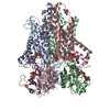
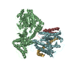
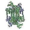
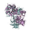
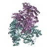
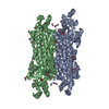
- PDB-6y9b: Cryo-EM structure of trimeric human STEAP1 bound to three Fab120.... -
+ Open data

- Basic information
| Entry | Database: PDB / ID: 6y9b | ||||||
|---|---|---|---|---|---|---|---|
| Title | Cryo-EM structure of trimeric human STEAP1 bound to three Fab120.545 fragments | ||||||
 Components Components |
| ||||||
 Keywords Keywords |  MEMBRANE PROTEIN / Integral membrane protein Heme-binding protein Oxidoreductase Antibody-antigen complex MEMBRANE PROTEIN / Integral membrane protein Heme-binding protein Oxidoreductase Antibody-antigen complex | ||||||
| Function / homology |  Function and homology information Function and homology informationcell-cell junction / endosome membrane / endosome / heme binding / membrane / metal ion binding / plasma membraneSimilarity search - Function | ||||||
| Biological species |  Homo sapiens (human) Homo sapiens (human) Mus musculus (house mouse) Mus musculus (house mouse) | ||||||
| Method | ELECTRON MICROSCOPY / single particle reconstruction / cryo EM / Resolution: 2.97 Å | ||||||
 Authors Authors | Oosterheert, W. / Gros, P. | ||||||
| Funding support |  Netherlands, 1items Netherlands, 1items
| ||||||
 Citation Citation | Journal: J Biol Chem / Year: 2020 Title: Cryo-electron microscopy structure and potential enzymatic function of human six-transmembrane epithelial antigen of the prostate 1 (STEAP1). Authors: Wout Oosterheert / Piet Gros / Abstract: Six-transmembrane epithelial antigen of the prostate 1 (STEAP1) is an integral membrane protein that is highly up-regulated on the cell surface of several human cancers, making it a promising ...Six-transmembrane epithelial antigen of the prostate 1 (STEAP1) is an integral membrane protein that is highly up-regulated on the cell surface of several human cancers, making it a promising therapeutic target to manage these diseases. It shares sequence homology with three enzymes (STEAP2-STEAP4) that catalyze the NADPH-dependent reduction of iron(III). However, STEAP1 lacks an intracellular NADPH-binding domain and does not exhibit cellular ferric reductase activity. Thus, both the molecular function of STEAP1 and its role in cancer progression remain elusive. Here, we present a ∼3.0-Å cryo-EM structure of trimeric human STEAP1 bound to three antigen-binding fragments (Fabs) of the clinically used antibody mAb120.545. The structure revealed that STEAP1 adopts a reductase-like conformation and interacts with the Fabs through its extracellular helices. Enzymatic assays in human cells revealed that STEAP1 promotes iron(III) reduction when fused to the intracellular NADPH-binding domain of its family member STEAP4, suggesting that STEAP1 functions as a ferric reductase in STEAP heterotrimers. Our work provides a foundation for deciphering the molecular mechanisms of STEAP1 and may be useful in the design of new therapeutic strategies to target STEAP1 in cancer. | ||||||
| History |
|
- Structure visualization
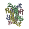
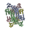
| Movie |
Movie viewer |
|---|---|
| Structure viewer | Molecule: MolmilJmol/JSmol |
- Downloads & links
-Download
| PDBx/mmCIF format | 6y9b.cif.gz | 281.7 KB | Display | PDBx/mmCIF format |
|---|---|---|---|---|
| PDB format | pdb6y9b.ent.gz | 226.9 KB | Display | PDB format |
| PDBx/mmJSON format | 6y9b.json.gz | Tree view | PDBx/mmJSON format | |
| Others |  Other downloads Other downloads |
-Validation report
| Arichive directory | https://data.pdbj.org/pub/pdb/validation_reports/y9/6y9bftp://data.pdbj.org/pub/pdb/validation_reports/y9/6y9b | HTTPS FTP |
|---|
-Related structure data
| Related structure data | 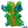 10735MC M: map data used to model this data C: citing same article ( |
|---|---|
| Similar structure data |

-Links
 PDBj
PDBj


- Assembly
| Deposited unit | 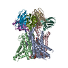
|
|---|---|
| 1 |
|
-Components
| #1: Protein | Mass: 44183.652 Da / Num. of mol.: 3 Source method: isolated from a genetically manipulated source Source: (gene. exp.) Homo sapiens (human) / Gene: STEAP1, PRSS24, STEAP / Plasmid: pUPE 2959Details (production host): Plasmid contains C-terminal Strep3-tag Cell line (production host): HEK293 GNT1- / Organ (production host): kidney / Production host: Homo sapiens (human) / Tissue (production host): embryonic kidneyReferences: UniProt: Q9UHE8, Oxidoreductases; Oxidizing metal ions; With NAD+ or NADP+ as acceptor#2: Antibody | Mass: 24344.135 Da / Num. of mol.: 3 Source method: isolated from a genetically manipulated source Details: Antibody fragment generated through mouse immunization Source: (gene. exp.) Mus musculus (house mouse) / Cell line (production host): HEK293 / Production host: Homo sapiens (human)#3: Antibody | Mass: 26149.922 Da / Num. of mol.: 3 Source method: isolated from a genetically manipulated source Details: Antibody fragment generated through mouse immunization Source: (gene. exp.) Mus musculus (house mouse) / Cell line (production host): HEK293 / Production host: Homo sapiens (human)#4: Chemical | Heme B  Mass: 616.487 Da / Num. of mol.: 3 / Source method: obtained synthetically / Formula: C34H32FeN4O4 / Feature type: SUBJECT OF INVESTIGATION Mass: 616.487 Da / Num. of mol.: 3 / Source method: obtained synthetically / Formula: C34H32FeN4O4 / Feature type: SUBJECT OF INVESTIGATION#5: Chemical | 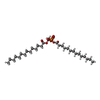  Mass: 591.777 Da / Num. of mol.: 3 / Source method: obtained synthetically / Formula: C31H60O8P / Comment: DMPA, phospholipid*YM Mass: 591.777 Da / Num. of mol.: 3 / Source method: obtained synthetically / Formula: C31H60O8P / Comment: DMPA, phospholipid*YMHas ligand of interest | Y | |
|---|
-Experimental details
-Experiment
| Experiment | Method: ELECTRON MICROSCOPY |
|---|---|
| EM experiment | Aggregation state: PARTICLE / 3D reconstruction method: single particle reconstruction |
- Sample preparation
| Component |
| ||||||||||||||||||||||||||||||||||||||||||
|---|---|---|---|---|---|---|---|---|---|---|---|---|---|---|---|---|---|---|---|---|---|---|---|---|---|---|---|---|---|---|---|---|---|---|---|---|---|---|---|---|---|---|---|
| Molecular weight |
| ||||||||||||||||||||||||||||||||||||||||||
| Source (natural) |
| ||||||||||||||||||||||||||||||||||||||||||
| Source (recombinant) |
| ||||||||||||||||||||||||||||||||||||||||||
| Buffer solution | pH: 7.8 Details: Protein sample was subjected to size-exclusion chromatography in 20 mM Tris pH 7.8, 200 mM NaCl, 0.08% (w/v) digitonin. 1 mM FAD (final concentration) was added to the sample before vitrification. | ||||||||||||||||||||||||||||||||||||||||||
| Buffer component |
| ||||||||||||||||||||||||||||||||||||||||||
| Specimen | Conc.: 5 mg/ml / Embedding applied: NO / Shadowing applied: NO / Staining applied: NO / Vitrification applied: YES Details: STEAP1-Fab120.545 complex purified in digitonin. The sample was monodisperse. | ||||||||||||||||||||||||||||||||||||||||||
| Specimen support | Grid material: GOLD / Grid mesh size: 200 divisions/in. / Grid type: Quantifoil R1.2/1.3 | ||||||||||||||||||||||||||||||||||||||||||
| Vitrification | Instrument: FEI VITROBOT MARK IV / Cryogen name: ETHANE-PROPANE / Humidity: 100 % / Chamber temperature: 293 K / Details: blot 4 seconds, blotforce 0 |
- Electron microscopy imaging
| Experimental equipment |  Model: Talos Arctica / Image courtesy: FEI Company |
|---|---|
| Microscopy | Model: FEI TALOS ARCTICA Details: Data was collected on a 200 kV Talos Arctica, located at Utrecht University, the Netherlands. |
| Electron gun | Electron source: FIELD EMISSION GUN / Accelerating voltage: 200 kV / Illumination mode: FLOOD BEAM |
| Electron lens | Mode: BRIGHT FIELDBright-field microscopy / Nominal magnification: 130000 X / Nominal defocus max: 3000 nm / Nominal defocus min: 1000 nm / Calibrated defocus min: 400 nm / Calibrated defocus max: 3500 nm / Cs: 2.7 mm / C2 aperture diameter: 50 µm / Alignment procedure: COMA FREE |
| Specimen holder | Cryogen: NITROGEN |
| Image recording | Average exposure time: 6.5 sec. / Electron dose: 49.5 e/Å2 / Detector mode: SUPER-RESOLUTION / Film or detector model: GATAN K2 SUMMIT (4k x 4k) / Num. of grids imaged: 1 / Num. of real images: 5325 |
| EM imaging optics | Energyfilter name: GIF Quantum LS / Energyfilter slit width: 20 eV |
| Image scans | Movie frames/image: 26 / Used frames/image: 1-26 |
- Processing
| Software | Name: PHENIX / Version: dev_3689: / Classification: refinement | ||||||||||||||||||||||||||||||||||||
|---|---|---|---|---|---|---|---|---|---|---|---|---|---|---|---|---|---|---|---|---|---|---|---|---|---|---|---|---|---|---|---|---|---|---|---|---|---|
| EM software |
| ||||||||||||||||||||||||||||||||||||
| CTF correction | Type: PHASE FLIPPING AND AMPLITUDE CORRECTION | ||||||||||||||||||||||||||||||||||||
| Particle selection | Num. of particles selected: 616302 / Details: Picking was performed in Relion. | ||||||||||||||||||||||||||||||||||||
| Symmetry | Point symmetry: C3 (3 fold cyclic) | ||||||||||||||||||||||||||||||||||||
| 3D reconstruction | Resolution: 2.97 Å / Resolution method: FSC 0.143 CUT-OFF / Num. of particles: 169426 / Details: Final reconstruction was generated in Relion. / Symmetry type: POINT | ||||||||||||||||||||||||||||||||||||
| Atomic model building | B value: 100.9 / Protocol: RIGID BODY FIT / Space: REAL | ||||||||||||||||||||||||||||||||||||
| Atomic model building | PDB-ID: 6HCY Pdb chain-ID: A / Pdb chain residue range: 196-454 | ||||||||||||||||||||||||||||||||||||
| Refine LS restraints |
|