 Movie
Movie Controller

[English] 日本語
 Yorodumi
- PDB-6y7q: Crystal Structure of the N-terminal PAS domain from the hERG3 Pot... -
+ Open data

- Basic information
| Entry | Database: PDB / ID: 6y7q | |||||||||
|---|---|---|---|---|---|---|---|---|---|---|
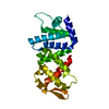
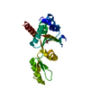
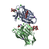
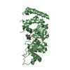
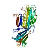
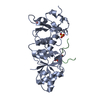
| Title | Crystal Structure of the N-terminal PAS domain from the hERG3 Potassium Channel | |||||||||
 Components Components | Potassium voltage-gated channel subfamily H member 7 | |||||||||
 Keywords Keywords |  MEMBRANE PROTEIN / PAS / Potassium Channel / Heme Binding / Rossmann Fold MEMBRANE PROTEIN / PAS / Potassium Channel / Heme Binding / Rossmann Fold | |||||||||
| Function / homology |  Function and homology information Function and homology informationVoltage gated Potassium channels / regulation of monoatomic ion transmembrane transport / voltage-gated potassium channel activity / plasma membrane => GO:0005886 / potassium ion transmembrane transport / regulation of membrane potential / plasma membraneSimilarity search - Function | |||||||||
| Biological species |  Homo sapiens (human) Homo sapiens (human) | |||||||||
| Method | X-RAY DIFFRACTION / SYNCHROTRON / MOLECULAR REPLACEMENT / Resolution: 1.39 Å | |||||||||
 Authors Authors | Cresser-Brown, J. / Raven, E. / Moody, P. | |||||||||
| Funding support |  United Kingdom, 2items United Kingdom, 2items
| |||||||||
 Citation Citation | Journal: J.Biol.Chem. / Year: 2020 Title: Discovery of a heme-binding domain in a neuronal voltage-gated potassium channel. Authors: Burton, M.J. / Cresser-Brown, J. / Thomas, M. / Portolano, N. / Basran, J. / Freeman, S.L. / Kwon, H. / Bottrill, A.R. / Llansola-Portoles, M.J. / Pascal, A.A. / Jukes-Jones, R. / Chernova, ...Authors: Burton, M.J. / Cresser-Brown, J. / Thomas, M. / Portolano, N. / Basran, J. / Freeman, S.L. / Kwon, H. / Bottrill, A.R. / Llansola-Portoles, M.J. / Pascal, A.A. / Jukes-Jones, R. / Chernova, T. / Schmid, R. / Davies, N.W. / Storey, N.M. / Dorlet, P. / Moody, P.C.E. / Mitcheson, J.S. / Raven, E.L. | |||||||||
| History |
|
- Structure visualization
| Structure viewer | Molecule: MolmilJmol/JSmol |
|---|
- Downloads & links
-Download
| PDBx/mmCIF format | 6y7q.cif.gz | 105.2 KB | Display | PDBx/mmCIF format |
|---|---|---|---|---|
| PDB format | pdb6y7q.ent.gz | Display | PDB format | |
| PDBx/mmJSON format | 6y7q.json.gz | Tree view | PDBx/mmJSON format | |
| Others |  Other downloads Other downloads |
-Validation report
| Arichive directory | https://data.pdbj.org/pub/pdb/validation_reports/y7/6y7qftp://data.pdbj.org/pub/pdb/validation_reports/y7/6y7q | HTTPS FTP |
|---|
-Related structure data
| Related structure data |  4hqaS S: Starting model for refinement |
|---|---|
| Similar structure data |


-Links
 PDBj
PDBj


- Assembly
| Deposited unit | 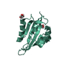
| ||||||||
|---|---|---|---|---|---|---|---|---|---|
| 1 | 
| ||||||||
| Unit cell |
|
-Components
| #1: Protein | Mass: 15392.727 Da / Num. of mol.: 1 Source method: isolated from a genetically manipulated source Source: (gene. exp.) Homo sapiens (human) / Gene: KCNH7, ERG3 / Plasmid: pLEICS-93 / Production host:  Escherichia coli BL21(DE3) (bacteria) / References: UniProt: Q9NS40 Escherichia coli BL21(DE3) (bacteria) / References: UniProt: Q9NS40 | ||||
|---|---|---|---|---|---|
| #2: Chemical | ChemComp-GOL / Glycerol  Mass: 92.094 Da / Num. of mol.: 1 / Source method: obtained synthetically / Formula: C3H8O3 Mass: 92.094 Da / Num. of mol.: 1 / Source method: obtained synthetically / Formula: C3H8O3 | ||||
| #3: Chemical | Ethylene glycol  Mass: 62.068 Da / Num. of mol.: 2 / Source method: obtained synthetically / Formula: C2H6O2 Mass: 62.068 Da / Num. of mol.: 2 / Source method: obtained synthetically / Formula: C2H6O2#4: Water | ChemComp-HOH / | Water Mass: 18.015 Da / Num. of mol.: 62 / Source method: isolated from a natural source / Formula: H2O Mass: 18.015 Da / Num. of mol.: 62 / Source method: isolated from a natural source / Formula: H2OHas ligand of interest | N | |
-Experimental details
-Experiment
| Experiment | Method: X-RAY DIFFRACTION / Number of used crystals: 1 |
|---|
- Sample preparation
| Crystal | Density Matthews: 1.75 Å3/Da / Density % sol: 29.78 % |
|---|---|
| Crystal grow | Temperature: 295 K / Method: vapor diffusion, sitting drop / pH: 7.5 Details: hERG3 PAS domain (15 mg/ml, in 50 mM NaCl, 1 mM TCEP in a 1:1 ratio with reservoir solution (0.1 M NaCl, 0.1 M HEPES pH=7.5, 1.6 M (NH4)2SO4 |
-Data collection
| Diffraction | Mean temperature: 100 K / Serial crystal experiment: N | ||||||||||||
|---|---|---|---|---|---|---|---|---|---|---|---|---|---|
| Diffraction source | Source: SYNCHROTRON / Site: Diamond / Beamline: I03 / Wavelength: 0.9795 Å | ||||||||||||
| Detector | Type: DECTRIS PILATUS3 6M / Detector: PIXEL / Date: Oct 30, 2017 | ||||||||||||
| Radiation | Protocol: SINGLE WAVELENGTH / Monochromatic (M) / Laue (L): M / Scattering type: x-ray | ||||||||||||
| Radiation wavelength | Wavelength: 0.9795 Å / Relative weight: 1 | ||||||||||||
| Reflection | Resolution: 1.39→66.9 Å / Num. obs: 23496 / % possible obs: 100 % / Redundancy: 11.9656537283 % / Rmerge(I) obs: 0.061 / Net I/σ(I): 13.9 | ||||||||||||
| Reflection shell |
|
- Processing
| Software |
| |||||||||||||||||||||||||||||||||||||||||||||||||||||||||||||||||||||||||||||||||||||||||||||||||||||||||||||||||||||||||||||||||||||||||||||||||||||||||||
|---|---|---|---|---|---|---|---|---|---|---|---|---|---|---|---|---|---|---|---|---|---|---|---|---|---|---|---|---|---|---|---|---|---|---|---|---|---|---|---|---|---|---|---|---|---|---|---|---|---|---|---|---|---|---|---|---|---|---|---|---|---|---|---|---|---|---|---|---|---|---|---|---|---|---|---|---|---|---|---|---|---|---|---|---|---|---|---|---|---|---|---|---|---|---|---|---|---|---|---|---|---|---|---|---|---|---|---|---|---|---|---|---|---|---|---|---|---|---|---|---|---|---|---|---|---|---|---|---|---|---|---|---|---|---|---|---|---|---|---|---|---|---|---|---|---|---|---|---|---|---|---|---|---|---|---|---|
| Refinement | Method to determine structure: MOLECULAR REPLACEMENT Starting model: 4HQA Resolution: 1.39→50.221 Å / Cor.coef. Fo:Fc: 0.972 / Cor.coef. Fo:Fc free: 0.96 / SU B: 7.176 / SU ML: 0.107 / Cross valid method: FREE R-VALUE / ESU R: 0.073 / ESU R Free: 0.071 Details: Hydrogens have been added in their riding positions
| |||||||||||||||||||||||||||||||||||||||||||||||||||||||||||||||||||||||||||||||||||||||||||||||||||||||||||||||||||||||||||||||||||||||||||||||||||||||||||
| Solvent computation | Ion probe radii: 0.8 Å / Shrinkage radii: 0.8 Å / VDW probe radii: 1.2 Å | |||||||||||||||||||||||||||||||||||||||||||||||||||||||||||||||||||||||||||||||||||||||||||||||||||||||||||||||||||||||||||||||||||||||||||||||||||||||||||
| Displacement parameters | Biso mean: 32.173 Å2
| |||||||||||||||||||||||||||||||||||||||||||||||||||||||||||||||||||||||||||||||||||||||||||||||||||||||||||||||||||||||||||||||||||||||||||||||||||||||||||
| Refinement step | Cycle: LAST / Resolution: 1.39→50.221 Å
| |||||||||||||||||||||||||||||||||||||||||||||||||||||||||||||||||||||||||||||||||||||||||||||||||||||||||||||||||||||||||||||||||||||||||||||||||||||||||||
| Refine LS restraints |
| |||||||||||||||||||||||||||||||||||||||||||||||||||||||||||||||||||||||||||||||||||||||||||||||||||||||||||||||||||||||||||||||||||||||||||||||||||||||||||
| LS refinement shell |
|
