 Movie
Movie Controller


+ Open data

- Basic information
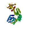
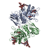
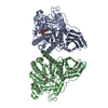
| Entry | Database: PDB / ID: 3mq1 | ||||||
|---|---|---|---|---|---|---|---|
| Title | Crystal Structure of Dust Mite Allergen Der p 5 | ||||||
 Components Components | Mite allergen Der p 5 | ||||||
 Keywords Keywords |  ALLERGEN / DUST MITE ALLERGEN / DUST MITE | ||||||
| Function / homology | Methane Monooxygenase Hydroxylase; Chain G, domain 1 - #970 / Mite allergen, group 5/21 / Mite allergen, group 5/21 superfamily / Mite allergen Blo t 5 / Methane Monooxygenase Hydroxylase; Chain G, domain 1 / Up-down Bundle / Mainly Alpha / PHOSPHATE ION / Mite allergen Der p 5 Function and homology information Function and homology information | ||||||
| Biological species |  Dermatophagoides pteronyssinus (European house dust mite) Dermatophagoides pteronyssinus (European house dust mite) | ||||||
| Method | X-RAY DIFFRACTION / Resolution: 2.8 Å | ||||||
 Authors Authors | Mueller, G.A. / Gosavi, R.A. / Krahn, J.M. / Edwards, L.L. / Cuneo, M.J. / Glesner, J. / Pomes, A. / Chapman, M.D. / London, R.E. / Pedersen, L.C. | ||||||
 Citation Citation | Journal: J.Biol.Chem. / Year: 2010 Title: Der p 5 crystal structure provides insight into the group 5 dust mite allergens. Authors: Mueller, G.A. / Gosavi, R.A. / Krahn, J.M. / Edwards, L.L. / Cuneo, M.J. / Glesner, J. / Pomes, A. / Chapman, M.D. / London, R.E. / Pedersen, L.C. | ||||||
| History |
|
- Structure visualization
| Structure viewer | Molecule: MolmilJmol/JSmol |
|---|
- Downloads & links
-Download
| PDBx/mmCIF format | 3mq1.cif.gz | 265.6 KB | Display | PDBx/mmCIF format |
|---|---|---|---|---|
| PDB format | pdb3mq1.ent.gz | 219.5 KB | Display | PDB format |
| PDBx/mmJSON format | 3mq1.json.gz | Tree view | PDBx/mmJSON format | |
| Others |  Other downloads Other downloads |
-Validation report
| Arichive directory | https://data.pdbj.org/pub/pdb/validation_reports/mq/3mq1ftp://data.pdbj.org/pub/pdb/validation_reports/mq/3mq1 | HTTPS FTP |
|---|
-Related structure data
| Similar structure data |
|---|


-Links
 PDBj
PDBj- Assembly
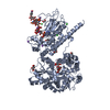
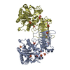
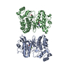
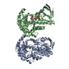
| Deposited unit | 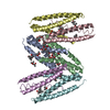
| ||||||||
|---|---|---|---|---|---|---|---|---|---|
| 1 | 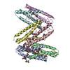
| ||||||||
| 2 | 
| ||||||||
| 3 | 
| ||||||||
| 4 | 
| ||||||||
| Unit cell |
|
-Components
| #1: Protein | Mass: 12143.111 Da / Num. of mol.: 6 / Fragment: UNP residues 34-132 Source method: isolated from a genetically manipulated source Source: (gene. exp.) Dermatophagoides pteronyssinus (European house dust mite)Gene: DERP5 / Plasmid: PGEX4T3(tev) / Production host:  Escherichia coli (E. coli) / Strain (production host): BL21(DE3)RIL / References: UniProt: P14004 Escherichia coli (E. coli) / Strain (production host): BL21(DE3)RIL / References: UniProt: P14004#2: Chemical | ChemComp-MPD / ( 2-Methyl-2,4-pentanediol  Mass: 118.174 Da / Num. of mol.: 17 / Source method: obtained synthetically / Formula: C6H14O2 / Comment: precipitant*YM Mass: 118.174 Da / Num. of mol.: 17 / Source method: obtained synthetically / Formula: C6H14O2 / Comment: precipitant*YM#3: Chemical | ChemComp-MRD / ( 2-Methyl-2,4-pentanediol  Mass: 118.174 Da / Num. of mol.: 17 / Source method: obtained synthetically / Formula: C6H14O2 / Comment: precipitant*YM Mass: 118.174 Da / Num. of mol.: 17 / Source method: obtained synthetically / Formula: C6H14O2 / Comment: precipitant*YM#4: Chemical | ChemComp-PO4 / | Phosphate  Mass: 94.971 Da / Num. of mol.: 1 / Source method: obtained synthetically / Formula: PO4 Mass: 94.971 Da / Num. of mol.: 1 / Source method: obtained synthetically / Formula: PO4#5: Water | ChemComp-HOH / | Water Mass: 18.015 Da / Num. of mol.: 45 / Source method: isolated from a natural source / Formula: H2O Mass: 18.015 Da / Num. of mol.: 45 / Source method: isolated from a natural source / Formula: H2O |
|---|
-Experimental details
-Experiment
| Experiment | Method: X-RAY DIFFRACTION / Number of used crystals: 1 |
|---|
- Sample preparation
| Crystal | Density Matthews: 2.6 Å3/Da / Density % sol: 52.74 % |
|---|---|
| Crystal grow | Temperature: 294 K / Method: vapor diffusion, sitting drop / pH: 6.2 Details: 31.5% MPD, 90 mM Na/K phosphate, pH 6.2, VAPOR DIFFUSION, SITTING DROP, temperature 294K |
-Data collection
| Diffraction | Mean temperature: 100 K |
|---|---|
| Diffraction source | Source: ROTATING ANODE / Type: RIGAKU MICROMAX-007 HF / Wavelength: 1.5418 Å |
| Detector | Type: RIGAKU SATURN 92 / Detector: CCD / Date: May 8, 2009 / Details: VariMax HF |
| Radiation | Monochromator: VariMaxHF mirrors / Protocol: SINGLE WAVELENGTH / Monochromatic (M) / Laue (L): M / Scattering type: x-ray |
| Radiation wavelength | Wavelength: 1.5418 Å / Relative weight: 1 |
| Reflection | Resolution: 2.8→50 Å / Num. obs: 19292 / % possible obs: 98.8 % / Observed criterion σ(F): 0 / Observed criterion σ(I): -3 / Redundancy: 6.4 % / Rsym value: 0.069 / Net I/σ(I): 15.7 |
| Reflection shell | Resolution: 2.8→2.9 Å / Redundancy: 4 % / Mean I/σ(I) obs: 2.5 / Num. unique all: 1883 / Rsym value: 0.518 / % possible all: 98.5 |
- Processing
| Software |
| ||||||||||||||||||||||||||||||||||||||||||||||||||||||||||||||||||||||||||||||||||||||||||||||||||||||||||||||||||||||||||||||||||||||||||||||||||||||||||||||||||||||||||||||||||||||
|---|---|---|---|---|---|---|---|---|---|---|---|---|---|---|---|---|---|---|---|---|---|---|---|---|---|---|---|---|---|---|---|---|---|---|---|---|---|---|---|---|---|---|---|---|---|---|---|---|---|---|---|---|---|---|---|---|---|---|---|---|---|---|---|---|---|---|---|---|---|---|---|---|---|---|---|---|---|---|---|---|---|---|---|---|---|---|---|---|---|---|---|---|---|---|---|---|---|---|---|---|---|---|---|---|---|---|---|---|---|---|---|---|---|---|---|---|---|---|---|---|---|---|---|---|---|---|---|---|---|---|---|---|---|---|---|---|---|---|---|---|---|---|---|---|---|---|---|---|---|---|---|---|---|---|---|---|---|---|---|---|---|---|---|---|---|---|---|---|---|---|---|---|---|---|---|---|---|---|---|---|---|---|---|
| Refinement | Resolution: 2.8→33.598 Å / SU ML: 0.38 / σ(F): 0.03 / Stereochemistry target values: ML
| ||||||||||||||||||||||||||||||||||||||||||||||||||||||||||||||||||||||||||||||||||||||||||||||||||||||||||||||||||||||||||||||||||||||||||||||||||||||||||||||||||||||||||||||||||||||
| Solvent computation | Shrinkage radii: 0.9 Å / VDW probe radii: 1.11 Å / Solvent model: FLAT BULK SOLVENT MODEL / Bsol: 67.539 Å2 / ksol: 0.287 e/Å3 | ||||||||||||||||||||||||||||||||||||||||||||||||||||||||||||||||||||||||||||||||||||||||||||||||||||||||||||||||||||||||||||||||||||||||||||||||||||||||||||||||||||||||||||||||||||||
| Refinement step | Cycle: LAST / Resolution: 2.8→33.598 Å
| ||||||||||||||||||||||||||||||||||||||||||||||||||||||||||||||||||||||||||||||||||||||||||||||||||||||||||||||||||||||||||||||||||||||||||||||||||||||||||||||||||||||||||||||||||||||
| Refine LS restraints |
| ||||||||||||||||||||||||||||||||||||||||||||||||||||||||||||||||||||||||||||||||||||||||||||||||||||||||||||||||||||||||||||||||||||||||||||||||||||||||||||||||||||||||||||||||||||||
| LS refinement shell |
| ||||||||||||||||||||||||||||||||||||||||||||||||||||||||||||||||||||||||||||||||||||||||||||||||||||||||||||||||||||||||||||||||||||||||||||||||||||||||||||||||||||||||||||||||||||||
| Refinement TLS params. | Refine-ID: X-RAY DIFFRACTION
| ||||||||||||||||||||||||||||||||||||||||||||||||||||||||||||||||||||||||||||||||||||||||||||||||||||||||||||||||||||||||||||||||||||||||||||||||||||||||||||||||||||||||||||||||||||||
| Refinement TLS group | Selection details: chain F |
