 Movie
Movie Controller


+ Open data

- Basic information
| Entry | Database: PDB / ID: 1r47 | |||||||||
|---|---|---|---|---|---|---|---|---|---|---|
| Title | Structure of human alpha-galactosidase | |||||||||
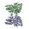
 Components Components | Alpha-galactosidase A | |||||||||
 Keywords Keywords |  HYDROLASE / glycoprotein / carbohydrate-binding protein / glycosidase / lysosomal enzyme / (beta/alpha)8 barrel HYDROLASE / glycoprotein / carbohydrate-binding protein / glycosidase / lysosomal enzyme / (beta/alpha)8 barrel | |||||||||
| Function / homology |  Function and homology information Function and homology informationglycosylceramide catabolic process / negative regulation of nitric-oxide synthase activity / alpha-galactosidase / alpha-galactosidase activity / glycosphingolipid catabolic process / galactoside binding / oligosaccharide metabolic process / glycoside catabolic process / negative regulation of nitric oxide biosynthetic process / Glycosphingolipid catabolism ...glycosylceramide catabolic process / negative regulation of nitric-oxide synthase activity / alpha-galactosidase / alpha-galactosidase activity / glycosphingolipid catabolic process / galactoside binding / oligosaccharide metabolic process / glycoside catabolic process / negative regulation of nitric oxide biosynthetic process / Glycosphingolipid catabolism / catalytic activity / lysosomal lumen / azurophil granule lumen / lysosome / hydrolase activity / signaling receptor binding / Neutrophil degranulation / Golgi apparatus / protein homodimerization activity / extracellular exosome / extracellular region / cytoplasmSimilarity search - Function | |||||||||
| Biological species |  Homo sapiens (human) Homo sapiens (human) | |||||||||
| Method | X-RAY DIFFRACTION / SYNCHROTRON / FOURIER SYNTHESIS / Resolution: 3.45 Å | |||||||||
 Authors Authors | Garman, S.C. / Garboczi, D.N. | |||||||||
 Citation Citation | Journal: J.Mol.Biol. / Year: 2004 Title: The molecular defect leading to Fabry disease: structure of human alpha-galactosidase Authors: Garman, S.C. / Garboczi, D.N. | |||||||||
| History |
|
- Structure visualization
| Structure viewer | Molecule: MolmilJmol/JSmol |
|---|
- Downloads & links
-Download
| PDBx/mmCIF format | 1r47.cif.gz | 177.5 KB | Display | PDBx/mmCIF format |
|---|---|---|---|---|
| PDB format | pdb1r47.ent.gz | 141.2 KB | Display | PDB format |
| PDBx/mmJSON format | 1r47.json.gz | Tree view | PDBx/mmJSON format | |
| Others |  Other downloads Other downloads |
-Validation report
| Arichive directory | https://data.pdbj.org/pub/pdb/validation_reports/r4/1r47ftp://data.pdbj.org/pub/pdb/validation_reports/r4/1r47 | HTTPS FTP |
|---|
-Related structure data


-Links
 PDBj
PDBj

- Assembly
| Deposited unit | 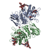
| ||||||||
|---|---|---|---|---|---|---|---|---|---|
| 1 |
| ||||||||
| Unit cell |
|
-Components
-Protein , 1 types, 2 molecules AB
| #1: Protein | Mass: 45394.543 Da / Num. of mol.: 2 Source method: isolated from a genetically manipulated source Details: complex with alpha-galactose / Source: (gene. exp.) Homo sapiens (human) / Gene: GLA / Production host: Homo sapiens (human) / References: UniProt: P06280, alpha-galactosidase |
|---|
-Sugars , 5 types, 8 molecules 
| #2: Polysaccharide | 2-acetamido-2-deoxy-beta-D-glucopyranose-(1-4)-2-acetamido-2-deoxy-beta-D-glucopyranose / Mass: 424.401 Da / Num. of mol.: 1 Source method: isolated from a genetically manipulated source | ||||||
|---|---|---|---|---|---|---|---|
| #3: Polysaccharide | / Mass: 910.823 Da / Num. of mol.: 2 Source method: isolated from a genetically manipulated source #4: Polysaccharide | / Mass: 586.542 Da / Num. of mol.: 2Source method: isolated from a genetically manipulated source #5: Polysaccharide | alpha-D-mannopyranose-(1-3)-[alpha-D-mannopyranose-(1-6)]alpha-D-mannopyranose-(1-4)-2-acetamido-2- ...alpha-D-mannopyranose-(1-3)-[alpha-D-mannopyranose-(1-6)]alpha-D-mannopyranose-(1-4)-2-acetamido-2-deoxy-beta-D-glucopyranose-(1-4)-[alpha-L-fucopyranose-(1-6)]2-acetamido-2-deoxy-beta-D-glucopyranose | / Mass: 1056.964 Da / Num. of mol.: 1Source method: isolated from a genetically manipulated source #6: Sugar | Galactose Type: D-saccharide, beta linking / Mass: 180.156 Da / Num. of mol.: 2 Type: D-saccharide, beta linking / Mass: 180.156 Da / Num. of mol.: 2Source method: isolated from a genetically manipulated source Formula: C6H12O6 |
-Non-polymers , 2 types, 20 molecules 

| #7: Chemical | Ethylene glycol Mass: 62.068 Da / Num. of mol.: 2 / Source method: obtained synthetically / Formula: C2H6O2 Mass: 62.068 Da / Num. of mol.: 2 / Source method: obtained synthetically / Formula: C2H6O2#8: Water | ChemComp-HOH / | WaterMass: 18.015 Da / Num. of mol.: 18 / Source method: isolated from a natural source / Formula: H2O |
|---|
-Experimental details
-Experiment
| Experiment | Method: X-RAY DIFFRACTION / Number of used crystals: 1 |
|---|
- Sample preparation
| Crystal | Density Matthews: 2.58 Å3/Da / Density % sol: 52 % | ||||||||||||||||||||||||||||||||||||
|---|---|---|---|---|---|---|---|---|---|---|---|---|---|---|---|---|---|---|---|---|---|---|---|---|---|---|---|---|---|---|---|---|---|---|---|---|---|
| Crystal grow | Temperature: 293 K / Method: vapor diffusion, sitting drop / pH: 8 Details: 30% PEG 4000, 0.1M TRIS HCl, 0.2M AMMONIUM SULFATE, pH 8.0, VAPOR DIFFUSION, SITTING DROP, temperature 293K | ||||||||||||||||||||||||||||||||||||
| Crystal grow | *PLUS pH: 7.5 / Method: vapor diffusion, hanging drop | ||||||||||||||||||||||||||||||||||||
| Components of the solutions | *PLUS
|
-Data collection
| Diffraction | Mean temperature: 100 K |
|---|---|
| Diffraction source | Source: SYNCHROTRON / Site: APS  / Beamline: 22-ID / Wavelength: 1.033 Å / Beamline: 22-ID / Wavelength: 1.033 Å |
| Detector | Type: MARRESEARCH / Detector: CCD / Date: Feb 23, 2003 |
| Radiation | Protocol: SINGLE WAVELENGTH / Monochromatic (M) / Laue (L): M / Scattering type: x-ray |
| Radiation wavelength | Wavelength: 1.033 Å / Relative weight: 1 |
| Reflection | Resolution: 3.45→41.55 Å / Num. all: 13878 / Num. obs: 13878 / % possible obs: 99.5 % / Observed criterion σ(F): 0 / Redundancy: 6.6 % / Biso Wilson estimate: 16 Å2 / Rmerge(I) obs: 0.2 / Rsym value: 0.2 / Net I/σ(I): 7.3 |
| Reflection shell | Resolution: 3.45→3.57 Å / Redundancy: 6.7 % / Rmerge(I) obs: 0.745 / Mean I/σ(I) obs: 2.4 / Num. unique all: 1323 / Rsym value: 0.745 / % possible all: 98.9 |
| Reflection | *PLUS Lowest resolution: 50 Å / Num. obs: 13922 / % possible obs: 99.7 % / Num. measured all: 91651 / Rmerge(I) obs: 0.2 |
| Reflection shell | *PLUS % possible obs: 98.9 % / Num. unique obs: 1323 / Num. measured obs: 8610 |
- Processing
| Software |
| ||||||||||||||||||||||||||||||||||||
|---|---|---|---|---|---|---|---|---|---|---|---|---|---|---|---|---|---|---|---|---|---|---|---|---|---|---|---|---|---|---|---|---|---|---|---|---|---|
| Refinement | Method to determine structure: FOURIER SYNTHESIS Starting model: Structure without ligand Resolution: 3.45→41.55 Å / Rfactor Rfree error: 0.012 / Isotropic thermal model: RESTRAINED / Cross valid method: THROUGHOUT / σ(F): 0 / Stereochemistry target values: Engh & Huber Details: 300 KCAL/MOL/A^2 NCS RESTRAINTS APPLIED TO ALL ATOMS IN EARLY ROUNDS OF REFINEMENT AND RELAXED IN LATER ROUNDS.
| ||||||||||||||||||||||||||||||||||||
| Solvent computation | Solvent model: FLAT MODEL / Bsol: 34.411 Å2 / ksol: 0.233224 e/Å3 | ||||||||||||||||||||||||||||||||||||
| Displacement parameters | Biso mean: 56.7 Å2
| ||||||||||||||||||||||||||||||||||||
| Refine analyze |
| ||||||||||||||||||||||||||||||||||||
| Refinement step | Cycle: LAST / Resolution: 3.45→41.55 Å
| ||||||||||||||||||||||||||||||||||||
| Refine LS restraints |
| ||||||||||||||||||||||||||||||||||||
| Refine LS restraints NCS | NCS model details: CONSTR / Weight Biso : 2 / Weight position: 300 | ||||||||||||||||||||||||||||||||||||
| LS refinement shell | Resolution: 3.45→3.67 Å / Rfactor Rfree error: 0.037 / Total num. of bins used: 6
| ||||||||||||||||||||||||||||||||||||
| Refinement | *PLUS Lowest resolution: 50 Å | ||||||||||||||||||||||||||||||||||||
| Solvent computation | *PLUS | ||||||||||||||||||||||||||||||||||||
| Displacement parameters | *PLUS | ||||||||||||||||||||||||||||||||||||
| Refine LS restraints | *PLUS
|
