 Movie
Movie Controller


+ Open data

- Basic information
| Entry | Database: PDB / ID: 6y7j | ||||||
|---|---|---|---|---|---|---|---|
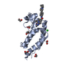
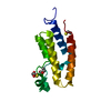
| Title | Structure of the BRD9 bromodomain and compound 15 | ||||||
 Components Components | Bromodomain-containing protein 9 | ||||||
 Keywords Keywords |  STRUCTURAL GENOMICS / BRD9 / Bromodomain-containing protein 9 STRUCTURAL GENOMICS / BRD9 / Bromodomain-containing protein 9 | ||||||
| Function / homology |  Function and homology information Function and homology informationGBAF complex / SWI/SNF complex / positive regulation of stem cell population maintenance / negative regulation of cell differentiation / lysine-acetylated histone binding / nucleic acid binding / chromatin remodeling / chromatin / positive regulation of cell population proliferation / regulation of transcription by RNA polymerase II ...GBAF complex / SWI/SNF complex / positive regulation of stem cell population maintenance / negative regulation of cell differentiation / lysine-acetylated histone binding / nucleic acid binding / chromatin remodeling / chromatin / positive regulation of cell population proliferation / regulation of transcription by RNA polymerase II / nucleoplasm / nucleusSimilarity search - Function | ||||||
| Biological species |  Homo sapiens (human) Homo sapiens (human) | ||||||
| Method | X-RAY DIFFRACTION / SYNCHROTRON / MOLECULAR REPLACEMENT / molecular replacement / Resolution: 1.6 Å | ||||||
 Authors Authors | Diaz-Saez, L. / Krojer, T. / Picaud, S. / von Delft, F. / Filippakopoulos, P. / Arrowsmith, C.H. / Edwards, A. / Bountra, C. / Huber, K.V.M. | ||||||
| Funding support | 1items
| ||||||
 Citation Citation | Journal: To Be Published Title: Structure of the BRD9 bromodomain Authors: Diaz-Saez, L. / Krojer, T. / Picaud, S. / von Delft, F. / Filippakopoulos, P. / Arrowsmith, C.H. / Edwards, A. / Bountra, C. / Huber, K.V.M. | ||||||
| History |
|
- Structure visualization
| Structure viewer | Molecule: MolmilJmol/JSmol |
|---|
- Downloads & links
-Download
| PDBx/mmCIF format | 6y7j.cif.gz | 69 KB | Display | PDBx/mmCIF format |
|---|---|---|---|---|
| PDB format | pdb6y7j.ent.gz | 49.9 KB | Display | PDB format |
| PDBx/mmJSON format | 6y7j.json.gz | Tree view | PDBx/mmJSON format | |
| Others |  Other downloads Other downloads |
-Validation report
| Arichive directory | https://data.pdbj.org/pub/pdb/validation_reports/y7/6y7jftp://data.pdbj.org/pub/pdb/validation_reports/y7/6y7j | HTTPS FTP |
|---|
-Related structure data
| Related structure data |  6y7hC  6y7kC  5mq1S S: Starting model for refinement C: citing same article ( |
|---|---|
| Similar structure data |


-Links
 PDBj
PDBj- Assembly
| Deposited unit | 
| |||||||||||||||||||||||||||
|---|---|---|---|---|---|---|---|---|---|---|---|---|---|---|---|---|---|---|---|---|---|---|---|---|---|---|---|---|
| 1 | 
| |||||||||||||||||||||||||||
| 2 | 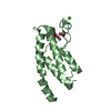
| |||||||||||||||||||||||||||
| Unit cell |
| |||||||||||||||||||||||||||
| Noncrystallographic symmetry (NCS) | NCS domain:
NCS domain segments:
|
-Components
| #1: Protein | Mass: 14031.487 Da / Num. of mol.: 2 Source method: isolated from a genetically manipulated source Source: (gene. exp.) Homo sapiens (human) / Gene: BRD9, UNQ3040/PRO9856 / Production host:  Escherichia coli BL21(DE3) (bacteria) / Variant (production host): R2 / References: UniProt: Q9H8M2 Escherichia coli BL21(DE3) (bacteria) / Variant (production host): R2 / References: UniProt: Q9H8M2#2: Chemical |   Mass: 296.321 Da / Num. of mol.: 2 / Source method: obtained synthetically / Formula: C17H16N2O3 / Feature type: SUBJECT OF INVESTIGATION Mass: 296.321 Da / Num. of mol.: 2 / Source method: obtained synthetically / Formula: C17H16N2O3 / Feature type: SUBJECT OF INVESTIGATION#3: Chemical | ChemComp-ACT / | Acetate  Mass: 59.044 Da / Num. of mol.: 1 / Source method: obtained synthetically / Formula: C2H3O2 Mass: 59.044 Da / Num. of mol.: 1 / Source method: obtained synthetically / Formula: C2H3O2#4: Chemical | ChemComp-CL / | Chloride  Mass: 35.453 Da / Num. of mol.: 1 / Source method: obtained synthetically / Formula: Cl Mass: 35.453 Da / Num. of mol.: 1 / Source method: obtained synthetically / Formula: Cl#5: Water | ChemComp-HOH / | Water Mass: 18.015 Da / Num. of mol.: 269 / Source method: isolated from a natural source / Formula: H2O Mass: 18.015 Da / Num. of mol.: 269 / Source method: isolated from a natural source / Formula: H2OHas ligand of interest | Y | |
|---|
-Experimental details
-Experiment
| Experiment | Method: X-RAY DIFFRACTION / Number of used crystals: 1 |
|---|
- Sample preparation
| Crystal | Density Matthews: 2.22 Å3/Da / Density % sol: 44.59 % |
|---|---|
| Crystal grow | Temperature: 277 K / Method: vapor diffusion, sitting drop / pH: 7.5 Details: 0.2M ammonium acetate, 25% PEG3350, 0.1M HEPES pH 7.5 |
-Data collection
| Diffraction | Mean temperature: 80 K / Serial crystal experiment: N | |||||||||||||||||||||||||||
|---|---|---|---|---|---|---|---|---|---|---|---|---|---|---|---|---|---|---|---|---|---|---|---|---|---|---|---|---|
| Diffraction source | Source: SYNCHROTRON / Site: Diamond  / Beamline: I04-1 / Wavelength: 0.92819 Å / Beamline: I04-1 / Wavelength: 0.92819 Å | |||||||||||||||||||||||||||
| Detector | Type: DECTRIS PILATUS 6M / Detector: PIXEL / Date: Sep 12, 2016 | |||||||||||||||||||||||||||
| Radiation | Protocol: SINGLE WAVELENGTH / Monochromatic (M) / Laue (L): M / Scattering type: x-ray | |||||||||||||||||||||||||||
| Radiation wavelength | Wavelength: 0.92819 Å / Relative weight: 1 | |||||||||||||||||||||||||||
| Reflection | Resolution: 1.6→62.86 Å / Num. obs: 34080 / % possible obs: 100 % / Redundancy: 5.9 % / CC1/2: 0.999 / Rmerge(I) obs: 0.073 / Rpim(I) all: 0.033 / Rrim(I) all: 0.081 / Net I/σ(I): 12 / Num. measured all: 202571 / Scaling rejects: 18 | |||||||||||||||||||||||||||
| Reflection shell | Diffraction-ID: 1 / Redundancy: 5.5 %
|
-Phasing
| Phasing | Method: molecular replacement | |||||||||
|---|---|---|---|---|---|---|---|---|---|---|
| Phasing MR | Model details: Phaser MODE: MR_AUTO
|
- Processing
| Software |
| ||||||||||||||||||||||||||||||||||||||||||||||||||||||||||||
|---|---|---|---|---|---|---|---|---|---|---|---|---|---|---|---|---|---|---|---|---|---|---|---|---|---|---|---|---|---|---|---|---|---|---|---|---|---|---|---|---|---|---|---|---|---|---|---|---|---|---|---|---|---|---|---|---|---|---|---|---|---|
| Refinement | Method to determine structure: MOLECULAR REPLACEMENT Starting model: 5MQ1 Resolution: 1.6→62.86 Å / Cor.coef. Fo:Fc: 0.963 / Cor.coef. Fo:Fc free: 0.941 / SU B: 2.466 / SU ML: 0.082 / Cross valid method: THROUGHOUT / σ(F): 0 / ESU R: 0.096 / ESU R Free: 0.103 Details: HYDROGENS HAVE BEEN ADDED IN THE RIDING POSITIONS U VALUES : REFINED INDIVIDUALLY
| ||||||||||||||||||||||||||||||||||||||||||||||||||||||||||||
| Solvent computation | Ion probe radii: 0.8 Å / Shrinkage radii: 0.8 Å / VDW probe radii: 1.2 Å | ||||||||||||||||||||||||||||||||||||||||||||||||||||||||||||
| Displacement parameters | Biso max: 92 Å2 / Biso mean: 28.47 Å2 / Biso min: 11.57 Å2
| ||||||||||||||||||||||||||||||||||||||||||||||||||||||||||||
| Refinement step | Cycle: final / Resolution: 1.6→62.86 Å
| ||||||||||||||||||||||||||||||||||||||||||||||||||||||||||||
| Refine LS restraints |
| ||||||||||||||||||||||||||||||||||||||||||||||||||||||||||||
| Refine LS restraints NCS | Ens-ID: 1 / Number: 6802 / Refine-ID: X-RAY DIFFRACTION / Type: interatomic distance / Rms dev position: 0.12 Å / Weight position: 0.05
| ||||||||||||||||||||||||||||||||||||||||||||||||||||||||||||
| LS refinement shell | Resolution: 1.6→1.642 Å / Rfactor Rfree error: 0 / Total num. of bins used: 20
|
