 Movie
Movie Controller


+ Open data

- Basic information
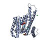
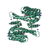
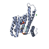
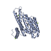
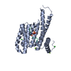
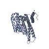
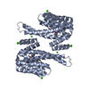
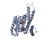
| Entry | Database: PDB / ID: 6y40 | ||||||
|---|---|---|---|---|---|---|---|
| Title | 14-3-3 Sigma in complex with phosphorylated PLN peptide | ||||||
 Components Components |
| ||||||
 Keywords Keywords |  PROTEIN BINDING / 14-3-3 / PLN / complex / protein / protein-protein interactions PROTEIN BINDING / 14-3-3 / PLN / complex / protein / protein-protein interactions | ||||||
| Function / homology |  Function and homology information Function and homology informationregulation of ATPase-coupled calcium transmembrane transporter activity / negative regulation of calcium ion binding / negative regulation of calcium ion import into sarcoplasmic reticulum / regulation of relaxation of cardiac muscle / negative regulation of ATPase-coupled calcium transmembrane transporter activity / adenylate cyclase-activating adrenergic receptor signaling pathway involved in heart process / regulation of the force of heart contraction by cardiac conduction / calcium ion-transporting ATPase complex / regulation of cardiac muscle cell membrane potential / negative regulation of calcium ion transmembrane transporter activity ...regulation of ATPase-coupled calcium transmembrane transporter activity / negative regulation of calcium ion binding / negative regulation of calcium ion import into sarcoplasmic reticulum / regulation of relaxation of cardiac muscle / negative regulation of ATPase-coupled calcium transmembrane transporter activity / adenylate cyclase-activating adrenergic receptor signaling pathway involved in heart process / regulation of the force of heart contraction by cardiac conduction / calcium ion-transporting ATPase complex / regulation of cardiac muscle cell membrane potential / negative regulation of calcium ion transmembrane transporter activity / acrosome assembly / negative regulation of calcium ion import / negative regulation of catalytic activity / ATPase inhibitor activity / regulation of the force of heart contraction / negative regulation of ATP-dependent activity / cardiac muscle tissue development / regulation of cardiac muscle cell contraction / blood circulation / enzyme inhibitor activity / regulation of heart contraction / relaxation of cardiac muscle / negative regulation of heart rate / muscle cell cellular homeostasis / regulation of epidermal cell division / protein kinase C inhibitor activity / positive regulation of epidermal cell differentiation / keratinocyte development / keratinization / regulation of calcium ion transport / Ion transport by P-type ATPases / negative regulation of calcium ion transport / Regulation of localization of FOXO transcription factors / keratinocyte proliferation / phosphoserine residue binding / Activation of BAD and translocation to mitochondria / negative regulation of keratinocyte proliferation / establishment of skin barrier / regulation of cardiac muscle contraction by regulation of the release of sequestered calcium ion / SARS-CoV-2 targets host intracellular signalling and regulatory pathways / Chk1/Chk2(Cds1) mediated inactivation of Cyclin B:Cdk1 complex / negative regulation of stem cell proliferation / SARS-CoV-1 targets host intracellular signalling and regulatory pathways / RHO GTPases activate PKNs / Ion homeostasis / Notch signaling pathway / protein kinase A signaling / sarcoplasmic reticulum membrane / regulation of cytosolic calcium ion concentration / protein export from nucleus / negative regulation of innate immune response / protein sequestering activity / TP53 Regulates Transcription of Genes Involved in G2 Cell Cycle Arrest / release of cytochrome c from mitochondria / positive regulation of protein export from nucleus / sarcoplasmic reticulum / stem cell proliferation / Translocation of SLC2A4 (GLUT4) to the plasma membrane / mitochondrial membrane / TP53 Regulates Metabolic Genes / negative regulation of protein kinase activity / negative regulation of cysteine-type endopeptidase activity involved in apoptotic process / intracellular calcium ion homeostasis / intrinsic apoptotic signaling pathway in response to DNA damage / ATPase binding / positive regulation of cell growth / regulation of cell cycle / cadherin binding / endoplasmic reticulum membrane / protein kinase binding / perinuclear region of cytoplasm / negative regulation of transcription by RNA polymerase II / endoplasmic reticulum / signal transduction / protein homodimerization activity / mitochondrion / extracellular space / extracellular exosome / membrane / identical protein binding / nucleus / cytosol / cytoplasmSimilarity search - Function | ||||||
| Biological species |  Homo sapiens (human) Homo sapiens (human) | ||||||
| Method | X-RAY DIFFRACTION / MOLECULAR REPLACEMENT / Resolution: 1.75 Å | ||||||
 Authors Authors | Ballone, A. / Lau, R.A. / Zweipfenning, F.P.A. / Ottmann, C. | ||||||
| Funding support |  Netherlands, 1items Netherlands, 1items
| ||||||
 Citation Citation | Journal: Acta Crystallogr.,Sect.F / Year: 2020 Title: A new soaking procedure for X-ray crystallographic structural determination of protein-peptide complexes. Authors: Ballone, A. / Lau, R.A. / Zweipfenning, F.P.A. / Ottmann, C. | ||||||
| History |
|
- Structure visualization
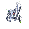
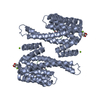
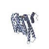
| Structure viewer | Molecule: MolmilJmol/JSmol |
|---|
- Downloads & links
-Download
| PDBx/mmCIF format | 6y40.cif.gz | 94 KB | Display | PDBx/mmCIF format |
|---|---|---|---|---|
| PDB format | pdb6y40.ent.gz | 55.9 KB | Display | PDB format |
| PDBx/mmJSON format | 6y40.json.gz | Tree view | PDBx/mmJSON format | |
| Others |  Other downloads Other downloads |
-Validation report
| Arichive directory | https://data.pdbj.org/pub/pdb/validation_reports/y4/6y40ftp://data.pdbj.org/pub/pdb/validation_reports/y4/6y40 | HTTPS FTP |
|---|
-Related structure data
| Related structure data |  6y3mC  6y3oC  6y3rC  6y3sC  6y3vC 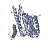 6y44C 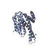 6y8aC 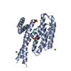 6y8bC 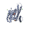 6y8dC 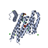 6y8eC 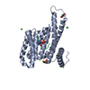 3lw1S S: Starting model for refinement C: citing same article ( |
|---|---|
| Similar structure data |


-Links
 PDBj
PDBj


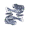
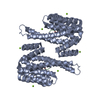
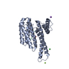
- Assembly
| Deposited unit | 
| ||||||||||||
|---|---|---|---|---|---|---|---|---|---|---|---|---|---|
| 1 | 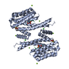
| ||||||||||||
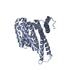
| Unit cell |
| ||||||||||||
| Components on special symmetry positions |
|
-Components
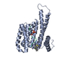
| #1: Protein/peptide | Mass: 1948.166 Da / Num. of mol.: 1 / Source method: obtained synthetically / Source: (synth.) Homo sapiens (human) / References: UniProt: P26678*PLUS | ||||||
|---|---|---|---|---|---|---|---|
| #2: Protein | Mass: 28226.518 Da / Num. of mol.: 1 Source method: isolated from a genetically manipulated source Source: (gene. exp.) Homo sapiens (human) / Gene: SFN, HME1 / Production host:  Escherichia coli (E. coli) / References: UniProt: P31947 Escherichia coli (E. coli) / References: UniProt: P31947 | ||||||
| #3: Chemical |   Mass: 24.305 Da / Num. of mol.: 3 / Source method: obtained synthetically / Formula: Mg Mass: 24.305 Da / Num. of mol.: 3 / Source method: obtained synthetically / Formula: Mg#4: Chemical | ChemComp-CL / | Chloride  Mass: 35.453 Da / Num. of mol.: 1 / Source method: obtained synthetically / Formula: Cl Mass: 35.453 Da / Num. of mol.: 1 / Source method: obtained synthetically / Formula: Cl#5: Water | ChemComp-HOH / | Water Mass: 18.015 Da / Num. of mol.: 517 / Source method: isolated from a natural source / Formula: H2O Mass: 18.015 Da / Num. of mol.: 517 / Source method: isolated from a natural source / Formula: H2OHas ligand of interest | Y | |
-Experimental details
-Experiment
| Experiment | Method: X-RAY DIFFRACTION / Number of used crystals: 1 |
|---|
- Sample preparation
| Crystal | Density Matthews: 2.4 Å3/Da / Density % sol: 48.7 % |
|---|---|
| Crystal grow | Temperature: 277 K / Method: vapor diffusion, hanging drop Details: PEG400, 1.25% glycerol, 0.2M CaCl, 0.1M HEPES pH 7.5, 2mM BME |
-Data collection
| Diffraction | Mean temperature: 100 K / Serial crystal experiment: N |
|---|---|
| Diffraction source | Source: SEALED TUBE / Type: RIGAKU / Wavelength: 1.541 Å |
| Detector | Type: DECTRIS PILATUS 200K / Detector: PIXEL / Date: Oct 16, 2019 |
| Radiation | Protocol: SINGLE WAVELENGTH / Monochromatic (M) / Laue (L): M / Scattering type: x-ray |
| Radiation wavelength | Wavelength: 1.541 Å / Relative weight: 1 |
| Reflection | Resolution: 1.59→41.81 Å / Num. obs: 31591 / % possible obs: 80.2 % / Biso Wilson estimate: 9.62 Å2 / CC1/2: 0.996 / Net I/σ(I): 14.3 |
| Reflection shell | Resolution: 1.59→1.63 Å / Num. unique obs: 31591 / CC1/2: 0.937 / % possible all: 98.8 |
- Processing
| Software |
| |||||||||||||||||||||||||||||||||||||||||||||||||||||||||||||||||||||||||||||
|---|---|---|---|---|---|---|---|---|---|---|---|---|---|---|---|---|---|---|---|---|---|---|---|---|---|---|---|---|---|---|---|---|---|---|---|---|---|---|---|---|---|---|---|---|---|---|---|---|---|---|---|---|---|---|---|---|---|---|---|---|---|---|---|---|---|---|---|---|---|---|---|---|---|---|---|---|---|---|
| Refinement | Method to determine structure: MOLECULAR REPLACEMENT Starting model: 3LW1 Resolution: 1.75→41.81 Å / SU ML: 0.1565 / Cross valid method: FREE R-VALUE / σ(F): 1.34 / Phase error: 19.4398
| |||||||||||||||||||||||||||||||||||||||||||||||||||||||||||||||||||||||||||||
| Solvent computation | Shrinkage radii: 0.9 Å / VDW probe radii: 1.11 Å | |||||||||||||||||||||||||||||||||||||||||||||||||||||||||||||||||||||||||||||
| Displacement parameters | Biso mean: 12.59 Å2 | |||||||||||||||||||||||||||||||||||||||||||||||||||||||||||||||||||||||||||||
| Refinement step | Cycle: LAST / Resolution: 1.75→41.81 Å
| |||||||||||||||||||||||||||||||||||||||||||||||||||||||||||||||||||||||||||||
| Refine LS restraints |
| |||||||||||||||||||||||||||||||||||||||||||||||||||||||||||||||||||||||||||||
| LS refinement shell |
|
