 Movie
Movie Controller

[English] 日本語
 Yorodumi
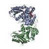
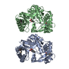
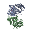
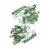
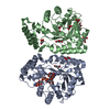
- PDB-6y1u: Mycobacterium tuberculosis FtsZ-GDP in complex with 4-hydroxycoumarin -
+ Open data

- Basic information
| Entry | Database: PDB / ID: 6y1u | ||||||
|---|---|---|---|---|---|---|---|
| Title | Mycobacterium tuberculosis FtsZ-GDP in complex with 4-hydroxycoumarin | ||||||
 Components Components | Cell division protein FtsZ | ||||||
 Keywords Keywords | ANTIBIOTIC / Cell division Protein | ||||||
| Function / homology |  Function and homology information Function and homology informationchloroplast fission / FtsZ-dependent cytokinesis / division septum assembly / cell division site / protein polymerization / GTPase activity / GTP binding / cytoplasmSimilarity search - Function | ||||||
| Biological species |   Mycobacterium tuberculosis (bacteria) Mycobacterium tuberculosis (bacteria) | ||||||
| Method | X-RAY DIFFRACTION / SYNCHROTRON / MOLECULAR REPLACEMENT / Resolution: 1.68 Å | ||||||
 Authors Authors | Alnami, A.T. / Norton, R.S. / Pena, H.P. / Haider, M. / kozielski, F. | ||||||
 Citation Citation | Journal: J.Mol.Biol. / Year: 2021 Title: Conformational Flexibility of A Highly Conserved Helix Controls Cryptic Pocket Formation in FtsZ. Authors: Alnami, A. / Norton, R.S. / Pena, H.P. / Haider, S. / Kozielski, F. | ||||||
| History |
|
- Structure visualization
| Structure viewer | Molecule: MolmilJmol/JSmol |
|---|
- Downloads & links
-Download
| PDBx/mmCIF format | 6y1u.cif.gz | 301.4 KB | Display | PDBx/mmCIF format |
|---|---|---|---|---|
| PDB format | pdb6y1u.ent.gz | 201.9 KB | Display | PDB format |
| PDBx/mmJSON format | 6y1u.json.gz | Tree view | PDBx/mmJSON format | |
| Others |  Other downloads Other downloads |
-Validation report
| Arichive directory | https://data.pdbj.org/pub/pdb/validation_reports/y1/6y1uftp://data.pdbj.org/pub/pdb/validation_reports/y1/6y1u | HTTPS FTP |
|---|
-Related structure data
| Related structure data | 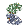 6y1vC 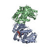 6ym1C  6ym9C  1rq7S S: Starting model for refinement C: citing same article ( |
|---|---|
| Similar structure data |


-Links
 PDBj
PDBj


- Assembly
| Deposited unit | 
| ||||||||||||
|---|---|---|---|---|---|---|---|---|---|---|---|---|---|
| 1 |
| ||||||||||||
| Unit cell |
|
-Components
| #1: Protein | Mass: 32083.266 Da / Num. of mol.: 2 Source method: isolated from a genetically manipulated source Source: (gene. exp.) Mycobacterium tuberculosis (strain CDC 1551 / Oshkosh) (bacteria)Strain: CDC 1551 / Oshkosh / Gene: ftsZ, MT2209 / Production host: Escherichia coli (E. coli) / References: UniProt: P9WN94#2: Chemical | ChemComp-DMS / Dimethyl sulfoxide  Mass: 78.133 Da / Num. of mol.: 21 / Source method: obtained synthetically / Formula: C2H6OS / Comment: DMSO, precipitant*YM Mass: 78.133 Da / Num. of mol.: 21 / Source method: obtained synthetically / Formula: C2H6OS / Comment: DMSO, precipitant*YM#3: Chemical | ChemComp-GDP / | Guanosine diphosphate  Type: RNA linking / Mass: 443.201 Da / Num. of mol.: 1 / Source method: obtained synthetically / Formula: C10H15N5O11P2 / Comment: GDP, energy-carrying molecule*YM Type: RNA linking / Mass: 443.201 Da / Num. of mol.: 1 / Source method: obtained synthetically / Formula: C10H15N5O11P2 / Comment: GDP, energy-carrying molecule*YM#4: Chemical | 4-Hydroxycoumarin  Mass: 162.142 Da / Num. of mol.: 2 / Source method: obtained synthetically / Formula: C9H6O3 Mass: 162.142 Da / Num. of mol.: 2 / Source method: obtained synthetically / Formula: C9H6O3#5: Water | ChemComp-HOH / | Water Mass: 18.015 Da / Num. of mol.: 396 / Source method: isolated from a natural source / Formula: H2O Mass: 18.015 Da / Num. of mol.: 396 / Source method: isolated from a natural source / Formula: H2OHas ligand of interest | Y | |
|---|
-Experimental details
-Experiment
| Experiment | Method: X-RAY DIFFRACTION / Number of used crystals: 1 |
|---|
- Sample preparation
| Crystal | Density Matthews: 3.07 Å3/Da / Density % sol: 59.99 % |
|---|---|
| Crystal grow | Temperature: 291.15 K / Method: vapor diffusion, hanging drop / pH: 5.6 Details: 27.5% PEG4000, 0.4 M ammonium acetate and 0.1 M Na citrate, soaked with 37.5 mM of inhibitor |
-Data collection
| Diffraction | Mean temperature: 298.15 K / Serial crystal experiment: N |
|---|---|
| Diffraction source | Source: SYNCHROTRON / Site: Diamond  / Beamline: I04-1 / Wavelength: 0.9159 Å / Beamline: I04-1 / Wavelength: 0.9159 Å |
| Detector | Type: DECTRIS PILATUS 6M-F / Detector: PIXEL / Date: Apr 5, 2019 |
| Radiation | Protocol: SINGLE WAVELENGTH / Monochromatic (M) / Laue (L): M / Scattering type: x-ray |
| Radiation wavelength | Wavelength: 0.9159 Å / Relative weight: 1 |
| Reflection | Resolution: 1.68→76.323 Å / Num. obs: 88000 / % possible obs: 100 % / Redundancy: 8 % / Biso Wilson estimate: 25.04 Å2 / CC1/2: 0.999 / Net I/σ(I): 17.5 |
| Reflection shell | Resolution: 1.68→1.74 Å / Num. unique obs: 8733 / CC1/2: 0.479 |
- Processing
| Software |
| |||||||||||||||||||||||||||||||||||||||||||||||||||||||||||||||||||||||||||||||||||||||||||||||||||||||||||||||||||||||||||||||||||||||||||||||||||||||||||||||||||||||||||||||||||||||||||||||||||||||||||||||||||||||||
|---|---|---|---|---|---|---|---|---|---|---|---|---|---|---|---|---|---|---|---|---|---|---|---|---|---|---|---|---|---|---|---|---|---|---|---|---|---|---|---|---|---|---|---|---|---|---|---|---|---|---|---|---|---|---|---|---|---|---|---|---|---|---|---|---|---|---|---|---|---|---|---|---|---|---|---|---|---|---|---|---|---|---|---|---|---|---|---|---|---|---|---|---|---|---|---|---|---|---|---|---|---|---|---|---|---|---|---|---|---|---|---|---|---|---|---|---|---|---|---|---|---|---|---|---|---|---|---|---|---|---|---|---|---|---|---|---|---|---|---|---|---|---|---|---|---|---|---|---|---|---|---|---|---|---|---|---|---|---|---|---|---|---|---|---|---|---|---|---|---|---|---|---|---|---|---|---|---|---|---|---|---|---|---|---|---|---|---|---|---|---|---|---|---|---|---|---|---|---|---|---|---|---|---|---|---|---|---|---|---|---|---|---|---|---|---|---|---|---|
| Refinement | Method to determine structure: MOLECULAR REPLACEMENT Starting model: 1RQ7 Resolution: 1.68→76.32 Å / SU ML: 0.1855 / Cross valid method: FREE R-VALUE / σ(F): 1.37 / Phase error: 20.2549 Stereochemistry target values: GeoStd + Monomer Library + CDL v1.2
| |||||||||||||||||||||||||||||||||||||||||||||||||||||||||||||||||||||||||||||||||||||||||||||||||||||||||||||||||||||||||||||||||||||||||||||||||||||||||||||||||||||||||||||||||||||||||||||||||||||||||||||||||||||||||
| Solvent computation | Shrinkage radii: 0.9 Å / VDW probe radii: 1.11 Å / Solvent model: FLAT BULK SOLVENT MODEL | |||||||||||||||||||||||||||||||||||||||||||||||||||||||||||||||||||||||||||||||||||||||||||||||||||||||||||||||||||||||||||||||||||||||||||||||||||||||||||||||||||||||||||||||||||||||||||||||||||||||||||||||||||||||||
| Displacement parameters | Biso mean: 33.9 Å2 | |||||||||||||||||||||||||||||||||||||||||||||||||||||||||||||||||||||||||||||||||||||||||||||||||||||||||||||||||||||||||||||||||||||||||||||||||||||||||||||||||||||||||||||||||||||||||||||||||||||||||||||||||||||||||
| Refinement step | Cycle: LAST / Resolution: 1.68→76.32 Å
| |||||||||||||||||||||||||||||||||||||||||||||||||||||||||||||||||||||||||||||||||||||||||||||||||||||||||||||||||||||||||||||||||||||||||||||||||||||||||||||||||||||||||||||||||||||||||||||||||||||||||||||||||||||||||
| Refine LS restraints |
| |||||||||||||||||||||||||||||||||||||||||||||||||||||||||||||||||||||||||||||||||||||||||||||||||||||||||||||||||||||||||||||||||||||||||||||||||||||||||||||||||||||||||||||||||||||||||||||||||||||||||||||||||||||||||
| LS refinement shell |
| |||||||||||||||||||||||||||||||||||||||||||||||||||||||||||||||||||||||||||||||||||||||||||||||||||||||||||||||||||||||||||||||||||||||||||||||||||||||||||||||||||||||||||||||||||||||||||||||||||||||||||||||||||||||||
| Refinement TLS params. | Method: refined / Origin x: -48.9133743045 Å / Origin y: 21.1360341601 Å / Origin z: 10.9634906097 Å
| |||||||||||||||||||||||||||||||||||||||||||||||||||||||||||||||||||||||||||||||||||||||||||||||||||||||||||||||||||||||||||||||||||||||||||||||||||||||||||||||||||||||||||||||||||||||||||||||||||||||||||||||||||||||||
| Refinement TLS group | Selection details: all |
