 Movie
Movie Controller

[English] 日本語
 Yorodumi
- PDB-6xy4: Structural insight into sheep-pox virus mediated inhibition of ap... -
+ Open data

- Basic information
| Entry | Database: PDB / ID: 6xy4 | |||||||||
|---|---|---|---|---|---|---|---|---|---|---|
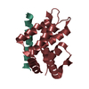
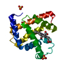
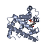
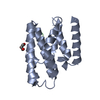
| Title | Structural insight into sheep-pox virus mediated inhibition of apoptosis | |||||||||
 Components Components |
| |||||||||
 Keywords Keywords |  APOPTOSIS / Pox virus / Bcl-2 APOPTOSIS / Pox virus / Bcl-2 | |||||||||
| Function / homology |  Function and homology information Function and homology information: / positive regulation of release of cytochrome c from mitochondria / positive regulation of protein-containing complex assembly / positive regulation of apoptotic process / apoptotic process / mitochondrion / membraneSimilarity search - Function | |||||||||
| Biological species | Sheeppox virus Homo sapiens (human) Homo sapiens (human) | |||||||||
| Method | X-RAY DIFFRACTION / SYNCHROTRON / MOLECULAR REPLACEMENT / Resolution: 2.04623155256 Å | |||||||||
 Authors Authors | Suraweera, C.D. / Hinds, M.G. / Kvansakul, M. | |||||||||
| Funding support |  Australia, 2items Australia, 2items
| |||||||||
 Citation Citation | Journal: Febs Lett. / Year: 2020 Title: Crystal structures of the sheeppox virus encoded inhibitor of apoptosis SPPV14 bound to the proapoptotic BH3 peptides Hrk and Bax. Authors: Suraweera, C.D. / Burton, D.R. / Hinds, M.G. / Kvansakul, M. | |||||||||
| History |
|
- Structure visualization
| Structure viewer | Molecule: MolmilJmol/JSmol |
|---|
- Downloads & links
-Download
| PDBx/mmCIF format | 6xy4.cif.gz | 122.8 KB | Display | PDBx/mmCIF format |
|---|---|---|---|---|
| PDB format | pdb6xy4.ent.gz | 80.4 KB | Display | PDB format |
| PDBx/mmJSON format | 6xy4.json.gz | Tree view | PDBx/mmJSON format | |
| Others |  Other downloads Other downloads |
-Validation report
| Arichive directory | https://data.pdbj.org/pub/pdb/validation_reports/xy/6xy4ftp://data.pdbj.org/pub/pdb/validation_reports/xy/6xy4 | HTTPS FTP |
|---|
-Related structure data
| Related structure data | 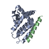 6xy6C 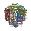 2bjyS S: Starting model for refinement C: citing same article ( |
|---|---|
| Similar structure data |


-Links
 PDBj
PDBj
- Assembly
| Deposited unit | 
| ||||||||||||
|---|---|---|---|---|---|---|---|---|---|---|---|---|---|
| 1 |
| ||||||||||||
| Unit cell |
|
-Components
| #1: Protein | Mass: 17476.027 Da / Num. of mol.: 1 Source method: isolated from a genetically manipulated source Source: (gene. exp.)  Sheeppox virus (strain Turkey/TU-V02127) Sheeppox virus (strain Turkey/TU-V02127)Gene: SPPV_14 / Production host:  Escherichia coli BL21(DE3) (bacteria) / Variant (production host): C41 / References: UniProt: A0A3F2YKH3 Escherichia coli BL21(DE3) (bacteria) / Variant (production host): C41 / References: UniProt: A0A3F2YKH3 |
|---|---|
| #2: Protein/peptide | Mass: 2973.417 Da / Num. of mol.: 1 / Source method: obtained synthetically / Source: (synth.) Homo sapiens (human) / References: UniProt: O00198 |
| #3: Water | ChemComp-HOH / Water Mass: 18.015 Da / Num. of mol.: 28 / Source method: isolated from a natural source / Formula: H2O Mass: 18.015 Da / Num. of mol.: 28 / Source method: isolated from a natural source / Formula: H2O |
-Experimental details
-Experiment
| Experiment | Method: X-RAY DIFFRACTION / Number of used crystals: 1 |
|---|
- Sample preparation
| Crystal | Density Matthews: 2.2 Å3/Da / Density % sol: 44.02 % |
|---|---|
| Crystal grow | Temperature: 293 K / Method: vapor diffusion, sitting drop / pH: 6.5 Details: 0.2 M Potassium sodium tartrate tetrahydrate, 0.1 M Bis-Tris propane pH 6.5, 20% w/vPEG 3350 |
-Data collection
| Diffraction | Mean temperature: 100 K / Serial crystal experiment: N |
|---|---|
| Diffraction source | Source: SYNCHROTRON / Site: Australian Synchrotron / Beamline: MX2 / Wavelength: 0.9537 Å |
| Detector | Type: DECTRIS EIGER X 16M / Detector: PIXEL / Date: Jul 18, 2019 |
| Radiation | Protocol: SINGLE WAVELENGTH / Monochromatic (M) / Laue (L): M / Scattering type: x-ray |
| Radiation wavelength | Wavelength: 0.9537 Å / Relative weight: 1 |
| Reflection | Resolution: 2.046→31.07 Å / Num. obs: 11236 / % possible obs: 98.2 % / Redundancy: 5 % / Biso Wilson estimate: 39.8950367928 Å2 / CC1/2: 0.99 / Rmerge(I) obs: 0.066 / Net I/σ(I): 8.7 |
| Reflection shell | Resolution: 2.05→8.92 Å / Num. unique obs: 782 / CC1/2: 0.573 |
- Processing
| Software |
| ||||||||||||||||||||||||||||||||||||||||
|---|---|---|---|---|---|---|---|---|---|---|---|---|---|---|---|---|---|---|---|---|---|---|---|---|---|---|---|---|---|---|---|---|---|---|---|---|---|---|---|---|---|
| Refinement | Method to determine structure: MOLECULAR REPLACEMENT Starting model: 2BJY Resolution: 2.04623155256→31.0657632133 Å / SU ML: 0.237702211781 / Cross valid method: FREE R-VALUE / σ(F): 1.38553218358 / Phase error: 26.211733487 Stereochemistry target values: GeoStd + Monomer Library + CDL v1.2
| ||||||||||||||||||||||||||||||||||||||||
| Solvent computation | Shrinkage radii: 0.9 Å / VDW probe radii: 1.11 Å / Solvent model: FLAT BULK SOLVENT MODEL | ||||||||||||||||||||||||||||||||||||||||
| Displacement parameters | Biso mean: 64.7478958779 Å2 | ||||||||||||||||||||||||||||||||||||||||
| Refinement step | Cycle: LAST / Resolution: 2.04623155256→31.0657632133 Å
| ||||||||||||||||||||||||||||||||||||||||
| Refine LS restraints |
| ||||||||||||||||||||||||||||||||||||||||
| LS refinement shell |
| ||||||||||||||||||||||||||||||||||||||||
| Refinement TLS params. | Method: refined / Origin x: -18.4590111381 Å / Origin y: -12.5771924003 Å / Origin z: -11.1701382726 Å
| ||||||||||||||||||||||||||||||||||||||||
| Refinement TLS group | Selection details: all |
