 Movie
Movie Controller

[English] 日本語
 Yorodumi
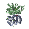
- PDB-6xut: Crystallographic structure of oligosaccharide dehydrogenase from ... -
+ Open data

- Basic information
| Entry | Database: PDB / ID: 6xut | |||||||||
|---|---|---|---|---|---|---|---|---|---|---|
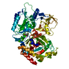
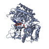
| Title | Crystallographic structure of oligosaccharide dehydrogenase from Pycnoporus cinnabarinus, ligand-free form | |||||||||
 Components Components | Oligosaccharide dehydrogenase | |||||||||
 Keywords Keywords |  OXIDOREDUCTASE / Glucose dehydrogenase / Pycnoporus cinnabarinus / oligosaccharide dehydrogenase OXIDOREDUCTASE / Glucose dehydrogenase / Pycnoporus cinnabarinus / oligosaccharide dehydrogenase | |||||||||
| Function / homology |  Function and homology information Function and homology informationoxidoreductase activity, acting on CH-OH group of donors / flavin adenine dinucleotide bindingSimilarity search - Function | |||||||||
| Biological species |  Pycnoporus cinnabarinus (fungus) Pycnoporus cinnabarinus (fungus) | |||||||||
| Method | X-RAY DIFFRACTION / SYNCHROTRON / MOLECULAR REPLACEMENT / Resolution: 1.6 Å | |||||||||
 Authors Authors | Cerutti, G. / Savino, C. / Montemiglio, L.C. / Sciara, G. / Vallone, B. | |||||||||
| Funding support |  France, 2items France, 2items
| |||||||||
 Citation Citation | Journal: Biotechnol Biofuels / Year: 2021 Title: Crystal structure and functional characterization of an oligosaccharide dehydrogenase from Pycnoporus cinnabarinus provides insights into fungal breakdown of lignocellulose. Authors: Cerutti, G. / Gugole, E. / Montemiglio, L.C. / Turbe-Doan, A. / Chena, D. / Navarro, D. / Lomascolo, A. / Piumi, F. / Exertier, C. / Freda, I. / Vallone, B. / Record, E. / Savino, C. / Sciara, G. | |||||||||
| History |
|
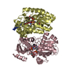
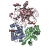
- Structure visualization
| Structure viewer | Molecule: MolmilJmol/JSmol |
|---|
- Downloads & links
-Download
| PDBx/mmCIF format | 6xut.cif.gz | 284.2 KB | Display | PDBx/mmCIF format |
|---|---|---|---|---|
| PDB format | pdb6xut.ent.gz | 228.3 KB | Display | PDB format |
| PDBx/mmJSON format | 6xut.json.gz | Tree view | PDBx/mmJSON format | |
| Others |  Other downloads Other downloads |
-Validation report
| Arichive directory | https://data.pdbj.org/pub/pdb/validation_reports/xu/6xutftp://data.pdbj.org/pub/pdb/validation_reports/xu/6xut | HTTPS FTP |
|---|
-Related structure data
| Related structure data | 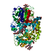 6xuuC 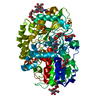 6xuvC 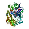 4yntS S: Starting model for refinement C: citing same article ( |
|---|---|
| Similar structure data |

-Links
 PDBj
PDBj
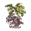
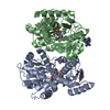
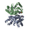
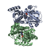
- Assembly
| Deposited unit | 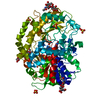
| ||||||||
|---|---|---|---|---|---|---|---|---|---|
| 1 |
| ||||||||
| Unit cell |
|
-Components
-Protein , 1 types, 1 molecules A
| #1: Protein | Mass: 62560.516 Da / Num. of mol.: 1 Source method: isolated from a genetically manipulated source Source: (gene. exp.) Pycnoporus cinnabarinus (fungus) / Gene: BN946_scf184803.g17 / Production host: Aspergillus niger (mold) / References: UniProt: A0A060SC37 |
|---|
-Sugars , 3 types, 3 molecules 
| #2: Polysaccharide | beta-D-mannopyranose-(1-3)-[beta-D-mannopyranose-(1-6)]beta-D-mannopyranose-(1-4)-2-acetamido-2- ...beta-D-mannopyranose-(1-3)-[beta-D-mannopyranose-(1-6)]beta-D-mannopyranose-(1-4)-2-acetamido-2-deoxy-beta-D-glucopyranose-(1-4)-2-acetamido-2-deoxy-beta-D-glucopyranose / Mass: 910.823 Da / Num. of mol.: 1 Source method: isolated from a genetically manipulated source |
|---|---|
| #3: Polysaccharide | 2-acetamido-2-deoxy-beta-D-glucopyranose-(1-4)-2-acetamido-2-deoxy-beta-D-glucopyranose / Mass: 424.401 Da / Num. of mol.: 1 / Source method: obtained synthetically |
| #5: Sugar | ChemComp-NAG / N-Acetylglucosamine Type: D-saccharide, beta linking / Mass: 221.208 Da / Num. of mol.: 1 / Source method: obtained synthetically / Formula: C8H15NO6 Type: D-saccharide, beta linking / Mass: 221.208 Da / Num. of mol.: 1 / Source method: obtained synthetically / Formula: C8H15NO6 |
-Non-polymers , 3 types, 463 molecules 


| #4: Chemical | ChemComp-FAD / Flavin adenine dinucleotide Mass: 785.550 Da / Num. of mol.: 1 / Source method: obtained synthetically / Formula: C27H33N9O15P2 / Feature type: SUBJECT OF INVESTIGATION / Comment: FAD*YM Mass: 785.550 Da / Num. of mol.: 1 / Source method: obtained synthetically / Formula: C27H33N9O15P2 / Feature type: SUBJECT OF INVESTIGATION / Comment: FAD*YM | ||
|---|---|---|---|
| #6: Chemical | ChemComp-SO4 / Sulfate Mass: 96.063 Da / Num. of mol.: 8 / Source method: isolated from a natural source / Formula: SO4 Mass: 96.063 Da / Num. of mol.: 8 / Source method: isolated from a natural source / Formula: SO4#7: Water | ChemComp-HOH / | WaterMass: 18.015 Da / Num. of mol.: 454 / Source method: isolated from a natural source / Formula: H2O |
-Details
| Has ligand of interest | Y |
|---|
-Experimental details
-Experiment
| Experiment | Method: X-RAY DIFFRACTION / Number of used crystals: 1 |
|---|
- Sample preparation
| Crystal | Density Matthews: 2.35 Å3/Da / Density % sol: 47.7 % |
|---|---|
| Crystal grow | Temperature: 294 K / Method: vapor diffusion, hanging drop / Details: 2 M ammonium sulfate |
-Data collection
| Diffraction | Mean temperature: 100 K / Serial crystal experiment: N |
|---|---|
| Diffraction source | Source: SYNCHROTRON / Site: Diamond  / Beamline: I24 / Wavelength: 1 Å / Beamline: I24 / Wavelength: 1 Å |
| Detector | Type: DECTRIS PILATUS3 6M / Detector: PIXEL / Date: Jun 28, 2019 |
| Radiation | Protocol: SINGLE WAVELENGTH / Monochromatic (M) / Laue (L): M / Scattering type: x-ray |
| Radiation wavelength | Wavelength: 1 Å / Relative weight: 1 |
| Reflection | Resolution: 1.43→97.74 Å / Num. obs: 103248 / % possible obs: 99.9 % / Redundancy: 12.7 % / CC1/2: 1 / Net I/σ(I): 14.5 |
| Reflection shell | Resolution: 1.43→1.57 Å / Mean I/σ(I) obs: 1.72 / Num. unique obs: 25836 / CC1/2: 0.79 |
- Processing
| Software |
| ||||||||||||||||||||||||||||||||||||||||||||||||||||||||||||||||||||||||||||||||||||||||||||||||||||||||||||||||||||||||||||||||||||||||||||||||||||||||||||||||||||||||||||||||||||||
|---|---|---|---|---|---|---|---|---|---|---|---|---|---|---|---|---|---|---|---|---|---|---|---|---|---|---|---|---|---|---|---|---|---|---|---|---|---|---|---|---|---|---|---|---|---|---|---|---|---|---|---|---|---|---|---|---|---|---|---|---|---|---|---|---|---|---|---|---|---|---|---|---|---|---|---|---|---|---|---|---|---|---|---|---|---|---|---|---|---|---|---|---|---|---|---|---|---|---|---|---|---|---|---|---|---|---|---|---|---|---|---|---|---|---|---|---|---|---|---|---|---|---|---|---|---|---|---|---|---|---|---|---|---|---|---|---|---|---|---|---|---|---|---|---|---|---|---|---|---|---|---|---|---|---|---|---|---|---|---|---|---|---|---|---|---|---|---|---|---|---|---|---|---|---|---|---|---|---|---|---|---|---|---|
| Refinement | Method to determine structure: MOLECULAR REPLACEMENT Starting model: 4YNT Resolution: 1.6→97.55 Å / Cor.coef. Fo:Fc: 0.975 / Cor.coef. Fo:Fc free: 0.965 / SU B: 6.123 / SU ML: 0.099 / Cross valid method: THROUGHOUT / ESU R: 0.089 / ESU R Free: 0.088 / Details: HYDROGENS HAVE BEEN ADDED IN THE RIDING POSITIONS
| ||||||||||||||||||||||||||||||||||||||||||||||||||||||||||||||||||||||||||||||||||||||||||||||||||||||||||||||||||||||||||||||||||||||||||||||||||||||||||||||||||||||||||||||||||||||
| Solvent computation | Ion probe radii: 0.8 Å / Shrinkage radii: 0.8 Å / VDW probe radii: 1.2 Å | ||||||||||||||||||||||||||||||||||||||||||||||||||||||||||||||||||||||||||||||||||||||||||||||||||||||||||||||||||||||||||||||||||||||||||||||||||||||||||||||||||||||||||||||||||||||
| Displacement parameters | Biso mean: 39.759 Å2
| ||||||||||||||||||||||||||||||||||||||||||||||||||||||||||||||||||||||||||||||||||||||||||||||||||||||||||||||||||||||||||||||||||||||||||||||||||||||||||||||||||||||||||||||||||||||
| Refinement step | Cycle: 1 / Resolution: 1.6→97.55 Å
| ||||||||||||||||||||||||||||||||||||||||||||||||||||||||||||||||||||||||||||||||||||||||||||||||||||||||||||||||||||||||||||||||||||||||||||||||||||||||||||||||||||||||||||||||||||||
| Refine LS restraints |
|