 Movie
Movie Controller

[English] 日本語
 Yorodumi
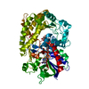
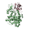
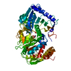
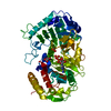
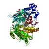
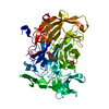
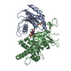
- PDB-4ynt: Crystal structure of Aspergillus flavus FAD glucose dehydrogenase -
+ Open data

- Basic information
| Entry | Database: PDB / ID: 4ynt | ||||||
|---|---|---|---|---|---|---|---|
| Title | Crystal structure of Aspergillus flavus FAD glucose dehydrogenase | ||||||
 Components Components | Glucose oxidase, putative | ||||||
 Keywords Keywords | OXIDOREDUCTASE / Glucose dehydrogenase / FAD | ||||||
| Function / homology |  Function and homology information Function and homology informationoxidoreductase activity, acting on CH-OH group of donors / flavin adenine dinucleotide bindingSimilarity search - Function | ||||||
| Biological species |  Aspergillus flavus NRRL3357 (mold) Aspergillus flavus NRRL3357 (mold) | ||||||
| Method | X-RAY DIFFRACTION / SYNCHROTRON / MOLECULAR REPLACEMENT / Resolution: 1.78 Å | ||||||
 Authors Authors | Yoshida, H. / Sakai, G. / Kojima, K. / Kamitori, S. / Sode, K. | ||||||
 Citation Citation | Journal: Sci Rep / Year: 2015 Title: Structural analysis of fungus-derived FAD glucose dehydrogenase Authors: Yoshida, H. / Sakai, G. / Mori, K. / Kojima, K. / Kamitori, S. / Sode, K. | ||||||
| History |
|
- Structure visualization
| Structure viewer | Molecule: MolmilJmol/JSmol |
|---|
- Downloads & links
-Download
| PDBx/mmCIF format | 4ynt.cif.gz | 134.9 KB | Display | PDBx/mmCIF format |
|---|---|---|---|---|
| PDB format | pdb4ynt.ent.gz | 101.6 KB | Display | PDB format |
| PDBx/mmJSON format | 4ynt.json.gz | Tree view | PDBx/mmJSON format | |
| Others |  Other downloads Other downloads |
-Validation report
| Arichive directory | https://data.pdbj.org/pub/pdb/validation_reports/yn/4yntftp://data.pdbj.org/pub/pdb/validation_reports/yn/4ynt | HTTPS FTP |
|---|
-Related structure data
| Related structure data |  4ynuC 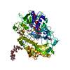 1cf3S C: citing same article ( S: Starting model for refinement |
|---|---|
| Similar structure data |


-Links
 PDBj
PDBj
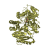
- Assembly
| Deposited unit | 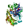
| ||||||||
|---|---|---|---|---|---|---|---|---|---|
| 1 |
| ||||||||
| Unit cell |
|
-Components
| #1: Protein | / glucose dehydrogenase Mass: 61548.922 Da / Num. of mol.: 1 / Fragment: UNP residues 24-593 Source method: isolated from a genetically manipulated source Source: (gene. exp.) Aspergillus flavus NRRL3357 (mold) / Strain: NRRL 3357 / Gene: AFLA_076820 / Plasmid: pET30c / Production host:  Escherichia coli (E. coli) / Strain (production host): Origami2(DE3) / References: UniProt: B8MX95, EC: 1.1.5.9 Escherichia coli (E. coli) / Strain (production host): Origami2(DE3) / References: UniProt: B8MX95, EC: 1.1.5.9 |
|---|---|
| #2: Chemical | ChemComp-FDA /   Mass: 787.566 Da / Num. of mol.: 1 / Source method: obtained synthetically / Formula: C27H35N9O15P2 Mass: 787.566 Da / Num. of mol.: 1 / Source method: obtained synthetically / Formula: C27H35N9O15P2 |
| #3: Water | ChemComp-HOH / Water Mass: 18.015 Da / Num. of mol.: 470 / Source method: isolated from a natural source / Formula: H2O Mass: 18.015 Da / Num. of mol.: 470 / Source method: isolated from a natural source / Formula: H2O |
-Experimental details
-Experiment
| Experiment | Method: X-RAY DIFFRACTION / Number of used crystals: 1 |
|---|
- Sample preparation
| Crystal | Density Matthews: 1.95 Å3/Da / Density % sol: 36.86 % |
|---|---|
| Crystal grow | Temperature: 293 K / Method: vapor diffusion, sitting drop / pH: 6.5 / Details: 0.1 M BisTris, 20-25 % PEG 3350 / PH range: 6.5 |
-Data collection
| Diffraction | Mean temperature: 100 K | |||||||||||||||||||||||||||||||||||||||||||||||||||||||||||||||||||||||||||||||||||||||||||||||||||||||||||||||||||||||||||||||||||||||||||||||||||
|---|---|---|---|---|---|---|---|---|---|---|---|---|---|---|---|---|---|---|---|---|---|---|---|---|---|---|---|---|---|---|---|---|---|---|---|---|---|---|---|---|---|---|---|---|---|---|---|---|---|---|---|---|---|---|---|---|---|---|---|---|---|---|---|---|---|---|---|---|---|---|---|---|---|---|---|---|---|---|---|---|---|---|---|---|---|---|---|---|---|---|---|---|---|---|---|---|---|---|---|---|---|---|---|---|---|---|---|---|---|---|---|---|---|---|---|---|---|---|---|---|---|---|---|---|---|---|---|---|---|---|---|---|---|---|---|---|---|---|---|---|---|---|---|---|---|---|---|---|
| Diffraction source | Source: SYNCHROTRON / Site: Photon Factory  / Beamline: BL-5A / Wavelength: 1 Å / Beamline: BL-5A / Wavelength: 1 Å | |||||||||||||||||||||||||||||||||||||||||||||||||||||||||||||||||||||||||||||||||||||||||||||||||||||||||||||||||||||||||||||||||||||||||||||||||||
| Detector | Type: ADSC QUANTUM 315r / Detector: CCD / Date: Jun 20, 2014 | |||||||||||||||||||||||||||||||||||||||||||||||||||||||||||||||||||||||||||||||||||||||||||||||||||||||||||||||||||||||||||||||||||||||||||||||||||
| Radiation | Protocol: SINGLE WAVELENGTH / Monochromatic (M) / Laue (L): M / Scattering type: x-ray | |||||||||||||||||||||||||||||||||||||||||||||||||||||||||||||||||||||||||||||||||||||||||||||||||||||||||||||||||||||||||||||||||||||||||||||||||||
| Radiation wavelength | Wavelength: 1 Å / Relative weight: 1 | |||||||||||||||||||||||||||||||||||||||||||||||||||||||||||||||||||||||||||||||||||||||||||||||||||||||||||||||||||||||||||||||||||||||||||||||||||
| Reflection | Resolution: 1.77→50 Å / Num. obs: 45130 / % possible obs: 99 % / Redundancy: 3.2 % / Rmerge(I) obs: 0.071 / Χ2: 1.381 / Net I/av σ(I): 17.424 / Net I/σ(I): 15.2 / Num. measured all: 143309 | |||||||||||||||||||||||||||||||||||||||||||||||||||||||||||||||||||||||||||||||||||||||||||||||||||||||||||||||||||||||||||||||||||||||||||||||||||
| Reflection shell | Diffraction-ID: 1 / Rejects: 0
|
- Processing
| Software |
| ||||||||||||||||||||||||||||||||||||||||||||||||||||||||||||
|---|---|---|---|---|---|---|---|---|---|---|---|---|---|---|---|---|---|---|---|---|---|---|---|---|---|---|---|---|---|---|---|---|---|---|---|---|---|---|---|---|---|---|---|---|---|---|---|---|---|---|---|---|---|---|---|---|---|---|---|---|---|
| Refinement | Method to determine structure: MOLECULAR REPLACEMENT Starting model: 1CF3 Resolution: 1.78→48.75 Å / Cor.coef. Fo:Fc: 0.969 / Cor.coef. Fo:Fc free: 0.954 / SU B: 2.602 / SU ML: 0.082 / Cross valid method: THROUGHOUT / σ(F): 0 / ESU R: 0.127 / ESU R Free: 0.122 / Stereochemistry target values: MAXIMUM LIKELIHOOD Details: HYDROGENS HAVE BEEN ADDED IN THE RIDING POSITIONS U VALUES : REFINED INDIVIDUALLY
| ||||||||||||||||||||||||||||||||||||||||||||||||||||||||||||
| Solvent computation | Ion probe radii: 0.8 Å / Shrinkage radii: 0.8 Å / VDW probe radii: 1.2 Å / Solvent model: MASK | ||||||||||||||||||||||||||||||||||||||||||||||||||||||||||||
| Displacement parameters | Biso max: 104.45 Å2 / Biso mean: 23.958 Å2 / Biso min: 11.82 Å2
| ||||||||||||||||||||||||||||||||||||||||||||||||||||||||||||
| Refinement step | Cycle: final / Resolution: 1.78→48.75 Å
| ||||||||||||||||||||||||||||||||||||||||||||||||||||||||||||
| Refine LS restraints |
| ||||||||||||||||||||||||||||||||||||||||||||||||||||||||||||
| LS refinement shell | Resolution: 1.775→1.821 Å / Total num. of bins used: 20
|
