 Movie
Movie Controller


+ Open data

- Basic information
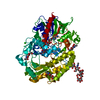
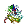
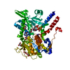
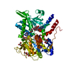
| Entry | Database: PDB / ID: 1cf3 | |||||||||
|---|---|---|---|---|---|---|---|---|---|---|
| Title | GLUCOSE OXIDASE FROM APERGILLUS NIGER | |||||||||
 Components Components | PROTEIN (GLUCOSE OXIDASE) | |||||||||
 Keywords Keywords | OXIDOREDUCTASE(FLAVOPROTEIN) | |||||||||
| Function / homology |  Function and homology information Function and homology information glucose oxidase / glucose oxidase activity / flavin adenine dinucleotide binding / extracellular region glucose oxidase / glucose oxidase activity / flavin adenine dinucleotide binding / extracellular regionSimilarity search - Function | |||||||||
| Biological species |  Aspergillus niger (mold) Aspergillus niger (mold) | |||||||||
| Method | X-RAY DIFFRACTION / SYNCHROTRON / MOLECULAR REPLACEMENT / Resolution: 1.9 Å | |||||||||
 Authors Authors | Hecht, H.J. / Kalisz, H. | |||||||||
 Citation Citation | Journal: Acta Crystallogr.,Sect.D / Year: 1999 Title: 1.8 and 1.9 A resolution structures of the Penicillium amagasakiense and Aspergillus niger glucose oxidases as a basis for modelling substrate complexes. Authors: Wohlfahrt, G. / Witt, S. / Hendle, J. / Schomburg, D. / Kalisz, H.M. / Hecht, H.J. #1: Journal: J.Mol.Biol. / Year: 1993Title: Crystal structure of glucose oxidase from Aspergillus niger refined at 2.3 A resolution. Authors: Hecht, H.J. / Kalisz, H.M. / Hendle, J. / Schmid, R.D. / Schomburg, D. #2: Journal: Biochim.Biophys.Acta / Year: 1991 Title: Effects of carbohydrate depletion on the structure, stability and activity of glucose oxidase from Aspergillus niger. Authors: Kalisz, H.M. / Hecht, H.J. / Schomburg, D. / Schmid, R.D. | |||||||||
| History |
|
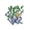
- Structure visualization
| Structure viewer | Molecule: MolmilJmol/JSmol |
|---|
- Downloads & links
-Download
| PDBx/mmCIF format | 1cf3.cif.gz | 136.3 KB | Display | PDBx/mmCIF format |
|---|---|---|---|---|
| PDB format | pdb1cf3.ent.gz | 103.7 KB | Display | PDB format |
| PDBx/mmJSON format | 1cf3.json.gz | Tree view | PDBx/mmJSON format | |
| Others |  Other downloads Other downloads |
-Validation report
| Arichive directory | https://data.pdbj.org/pub/pdb/validation_reports/cf/1cf3ftp://data.pdbj.org/pub/pdb/validation_reports/cf/1cf3 | HTTPS FTP |
|---|
-Related structure data
| Related structure data | 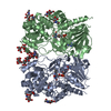 1gpeC  1galS S: Starting model for refinement C: citing same article ( |
|---|---|
| Similar structure data |


-Links
 PDBj
PDBj
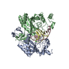
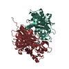
- Assembly
| Deposited unit | 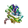
| ||||||||
|---|---|---|---|---|---|---|---|---|---|
| 1 |
| ||||||||
| Unit cell |
|
-Components
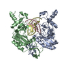
| #1: Protein | Mass: 63329.020 Da / Num. of mol.: 1 / Source method: isolated from a natural source / Source: (natural) Aspergillus niger (mold) / References: UniProt: P13006, glucose oxidase | ||||
|---|---|---|---|---|---|
| #2: Polysaccharide | alpha-D-mannopyranose-(1-2)-alpha-D-mannopyranose-(1-3)-beta-D-mannopyranose-(1-4)-2-acetamido-2- ...alpha-D-mannopyranose-(1-2)-alpha-D-mannopyranose-(1-3)-beta-D-mannopyranose-(1-4)-2-acetamido-2-deoxy-beta-D-glucopyranose-(1-4)-2-acetamido-2-deoxy-beta-D-glucopyranose / Mass: 910.823 Da / Num. of mol.: 1 Source method: isolated from a genetically manipulated source | ||||
| #3: Sugar | N-Acetylglucosamine  Type: D-saccharide, beta linking / Mass: 221.208 Da / Num. of mol.: 3 Type: D-saccharide, beta linking / Mass: 221.208 Da / Num. of mol.: 3Source method: isolated from a genetically manipulated source Formula: C8H15NO6 #4: Chemical | ChemComp-FAD / | Flavin adenine dinucleotide  Mass: 785.550 Da / Num. of mol.: 1 / Source method: obtained synthetically / Formula: C27H33N9O15P2 / Comment: FAD*YM Mass: 785.550 Da / Num. of mol.: 1 / Source method: obtained synthetically / Formula: C27H33N9O15P2 / Comment: FAD*YM#5: Water | ChemComp-HOH / | Water Mass: 18.015 Da / Num. of mol.: 309 / Source method: isolated from a natural source / Formula: H2O Mass: 18.015 Da / Num. of mol.: 309 / Source method: isolated from a natural source / Formula: H2O |
-Experimental details
-Experiment
| Experiment | Method: X-RAY DIFFRACTION / Number of used crystals: 1 |
|---|
- Sample preparation
| Crystal | Density Matthews: 2.2 Å3/Da / Density % sol: 43.2 % | ||||||||||||||||||||||||||||||||||||
|---|---|---|---|---|---|---|---|---|---|---|---|---|---|---|---|---|---|---|---|---|---|---|---|---|---|---|---|---|---|---|---|---|---|---|---|---|---|
| Crystal grow | pH: 5.6 / Details: pH 5.6 | ||||||||||||||||||||||||||||||||||||
| Crystal | *PLUS | ||||||||||||||||||||||||||||||||||||
| Crystal grow | *PLUS Method: vapor diffusion, hanging drop / Details: Kalisz, H.M., (1990) J.Mol.Biol., 213, 207. / PH range low: 5.6 / PH range high: 5.3 | ||||||||||||||||||||||||||||||||||||
| Components of the solutions | *PLUS
|
-Data collection
| Diffraction | Mean temperature: 290 K |
|---|---|
| Diffraction source | Source: SYNCHROTRON / Site: MPG/DESY, HAMBURG  / Beamline: BW6 / Wavelength: 1 / Beamline: BW6 / Wavelength: 1 |
| Detector | Type: MARRESEARCH / Detector: IMAGE PLATE / Details: MIRRORS |
| Radiation | Monochromator: SI(111) / Protocol: SINGLE WAVELENGTH / Monochromatic (M) / Laue (L): M / Scattering type: x-ray |
| Radiation wavelength | Wavelength: 1 Å / Relative weight: 1 |
| Reflection | Resolution: 1.9→20 Å / Num. obs: 40401 / % possible obs: 87.7 % / Observed criterion σ(I): 0 / Redundancy: 2.5 % / Biso Wilson estimate: 20.24 Å2 / Rmerge(I) obs: 0.054 / Net I/σ(I): 21.5 |
| Reflection shell | Resolution: 1.9→1.99 Å / Redundancy: 1.5 % / Rmerge(I) obs: 0.122 / Mean I/σ(I) obs: 6.9 / % possible all: 78.7 |
| Reflection shell | *PLUS Highest resolution: 1.9 Å / % possible obs: 78.7 % |
- Processing
| Software |
| ||||||||||||||||||||||||||||||||||||||||||||||||||||||||||||||||||||||||||||||||||||
|---|---|---|---|---|---|---|---|---|---|---|---|---|---|---|---|---|---|---|---|---|---|---|---|---|---|---|---|---|---|---|---|---|---|---|---|---|---|---|---|---|---|---|---|---|---|---|---|---|---|---|---|---|---|---|---|---|---|---|---|---|---|---|---|---|---|---|---|---|---|---|---|---|---|---|---|---|---|---|---|---|---|---|---|---|---|
| Refinement | Method to determine structure: MOLECULAR REPLACEMENT Starting model: PDB ENTRY 1GAL Resolution: 1.9→20 Å / Cross valid method: THROUGHOUT / σ(F): 0 / ESU R: 0.2
| ||||||||||||||||||||||||||||||||||||||||||||||||||||||||||||||||||||||||||||||||||||
| Displacement parameters | Biso mean: 24.22 Å2 | ||||||||||||||||||||||||||||||||||||||||||||||||||||||||||||||||||||||||||||||||||||
| Refinement step | Cycle: LAST / Resolution: 1.9→20 Å
| ||||||||||||||||||||||||||||||||||||||||||||||||||||||||||||||||||||||||||||||||||||
| Refine LS restraints |
| ||||||||||||||||||||||||||||||||||||||||||||||||||||||||||||||||||||||||||||||||||||
| Software | *PLUS Name: REFMAC / Classification: refinement | ||||||||||||||||||||||||||||||||||||||||||||||||||||||||||||||||||||||||||||||||||||
| Refinement | *PLUS Lowest resolution: 20 Å / Rfactor obs: 0.19 / Rfactor Rfree: 0.243 | ||||||||||||||||||||||||||||||||||||||||||||||||||||||||||||||||||||||||||||||||||||
| Solvent computation | *PLUS | ||||||||||||||||||||||||||||||||||||||||||||||||||||||||||||||||||||||||||||||||||||
| Displacement parameters | *PLUS | ||||||||||||||||||||||||||||||||||||||||||||||||||||||||||||||||||||||||||||||||||||
| Refine LS restraints | *PLUS
| ||||||||||||||||||||||||||||||||||||||||||||||||||||||||||||||||||||||||||||||||||||
| LS refinement shell | *PLUS Highest resolution: 1.9 Å / Lowest resolution: 1.99 Å / Rfactor Rfree: 0.329 / Rfactor obs: 0.242 |
