 Movie
Movie Controller


+ Open data

- Basic information
| Entry | Database: PDB / ID: 6xnm | |||||||||
|---|---|---|---|---|---|---|---|---|---|---|
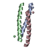
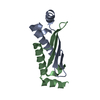
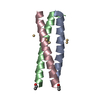
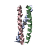
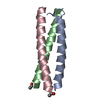
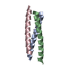
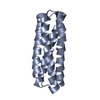
| Title | GCN4-p1 Peptide Trimer with tyrosine residue at position 16 | |||||||||
 Components Components |
| |||||||||
 Keywords Keywords |  TRANSCRIPTION / Coil-coil / tyrosine TRANSCRIPTION / Coil-coil / tyrosine | |||||||||
| Function / homology |  Function and homology information Function and homology informationprotein localization to nuclear periphery / Activation of the AP-1 family of transcription factors / response to amino acid starvation / mediator complex binding / negative regulation of ribosomal protein gene transcription by RNA polymerase II / positive regulation of cellular response to amino acid starvation / nitrogen catabolite activation of transcription from RNA polymerase II promoter / TFIID-class transcription factor complex binding / amino acid biosynthetic process / positive regulation of transcription initiation by RNA polymerase II ...protein localization to nuclear periphery / Activation of the AP-1 family of transcription factors / response to amino acid starvation / mediator complex binding / negative regulation of ribosomal protein gene transcription by RNA polymerase II / positive regulation of cellular response to amino acid starvation / nitrogen catabolite activation of transcription from RNA polymerase II promoter / TFIID-class transcription factor complex binding / amino acid biosynthetic process / positive regulation of transcription initiation by RNA polymerase II / positive regulation of RNA polymerase II transcription preinitiation complex assembly / cellular response to amino acid starvation / RNA polymerase II transcription regulator complex / : / DNA-binding transcription activator activity, RNA polymerase II-specific / transcription regulator complex / RNA polymerase II-specific DNA-binding transcription factor binding / sequence-specific DNA binding / DNA-binding transcription factor activity, RNA polymerase II-specific / intracellular signal transduction / RNA polymerase II cis-regulatory region sequence-specific DNA binding / DNA-binding transcription factor activity / chromatin binding / negative regulation of transcription by RNA polymerase II / positive regulation of transcription by RNA polymerase II / identical protein binding / nucleusSimilarity search - Function | |||||||||
| Biological species |  Saccharomyces cerevisiae (brewer's yeast) Saccharomyces cerevisiae (brewer's yeast) | |||||||||
| Method | X-RAY DIFFRACTION / MOLECULAR REPLACEMENT / Resolution: 2.25 Å | |||||||||
 Authors Authors | Rowe Hartje, R.K. / Czarny, R.S. / Ho, A. | |||||||||
| Funding support |  United States, 2items United States, 2items
| |||||||||
 Citation Citation | Journal: To Be Published Title: Engineering Specific Protein-Protein Interactions Through Halogen and Hydrogen Bonds Authors: Rowe Hartje, R.K. / Ferrero, M. / Cavallo, G. / Metrangolo, P. / Ho, A. / Czarny, R. / Ho, P.S. | |||||||||
| History |
|
- Structure visualization
| Structure viewer | Molecule: MolmilJmol/JSmol |
|---|
- Downloads & links
-Download
| PDBx/mmCIF format | 6xnm.cif.gz | 61.5 KB | Display | PDBx/mmCIF format |
|---|---|---|---|---|
| PDB format | pdb6xnm.ent.gz | 38.6 KB | Display | PDB format |
| PDBx/mmJSON format | 6xnm.json.gz | Tree view | PDBx/mmJSON format | |
| Others |  Other downloads Other downloads |
-Validation report
| Arichive directory | https://data.pdbj.org/pub/pdb/validation_reports/xn/6xnmftp://data.pdbj.org/pub/pdb/validation_reports/xn/6xnm | HTTPS FTP |
|---|
-Related structure data
| Related structure data |  6xneC 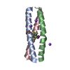 6xnfC  6xnlC  1swiS S: Starting model for refinement C: citing same article ( |
|---|---|
| Similar structure data |


-Links
 PDBj
PDBj

- Assembly
| Deposited unit | 
| ||||||||||||
|---|---|---|---|---|---|---|---|---|---|---|---|---|---|
| 1 |
| ||||||||||||
| Unit cell |
| ||||||||||||
| Components on special symmetry positions |
|
-Components
| #1: Protein/peptide | Mass: 3619.280 Da / Num. of mol.: 2 / Mutation: N16A / Source method: obtained synthetically / Source: (synth.) Saccharomyces cerevisiae (brewer's yeast) / References: UniProt: P03069#2: Protein/peptide | | Mass: 3711.375 Da / Num. of mol.: 1 / Mutation: N16Y / Source method: obtained synthetically / Source: (synth.) Saccharomyces cerevisiae (brewer's yeast) / References: UniProt: P03069#3: Chemical | ChemComp-NA /   Mass: 22.990 Da / Num. of mol.: 7 / Mutation: N16Y / Source method: obtained synthetically / Formula: Na Mass: 22.990 Da / Num. of mol.: 7 / Mutation: N16Y / Source method: obtained synthetically / Formula: Na#4: Water | ChemComp-HOH / | Water Mass: 18.015 Da / Num. of mol.: 43 / Source method: isolated from a natural source / Formula: H2O Mass: 18.015 Da / Num. of mol.: 43 / Source method: isolated from a natural source / Formula: H2OHas ligand of interest | N | |
|---|
-Experimental details
-Experiment
| Experiment | Method: X-RAY DIFFRACTION / Number of used crystals: 1 |
|---|
- Sample preparation
| Crystal | Density Matthews: 2.16 Å3/Da / Density % sol: 43.13 % |
|---|---|
| Crystal grow | Temperature: 298 K / Method: vapor diffusion, hanging drop / pH: 7 Details: Crystallization drops were prepared by mixing 2 uL of stock peptide solution with 2 uL of mother liquor and allowed to equilibrate at 298 K over a well containing 500 uL of mother liquor. ...Details: Crystallization drops were prepared by mixing 2 uL of stock peptide solution with 2 uL of mother liquor and allowed to equilibrate at 298 K over a well containing 500 uL of mother liquor. The stock peptide solution (total concentration 1.5 mM) was prepared by mixing 2:1 ratios of the A16 peptide with me-F16 in 10 mM potassium phosphate, 100 mM potassium chloride pH 7.0. |
-Data collection
| Diffraction | Mean temperature: 100 K / Serial crystal experiment: N |
|---|---|
| Diffraction source | Source: ROTATING ANODE / Type: RIGAKU MICROMAX-003 / Wavelength: 1.5418 Å |
| Detector | Type: DECTRIS PILATUS 200K / Detector: PIXEL / Date: Sep 1, 2017 |
| Radiation | Protocol: SINGLE WAVELENGTH / Monochromatic (M) / Laue (L): M / Scattering type: x-ray |
| Radiation wavelength | Wavelength: 1.5418 Å / Relative weight: 1 |
| Reflection | Resolution: 2.25→13.77 Å / Num. obs: 7950 / % possible obs: 91.53 % / Redundancy: 1.9 % / Biso Wilson estimate: 22.67 Å2 / CC1/2: 0.996 / CC star: 0.999 / Rmerge(I) obs: 0.04139 / Rpim(I) all: 0.04139 / Rrim(I) all: 0.05854 / Net I/σ(I): 20.01 |
| Reflection shell | Resolution: 2.25→2.33 Å / Redundancy: 1.9 % / Rmerge(I) obs: 0.08967 / Mean I/σ(I) obs: 9.6 / Num. unique obs: 818 / CC1/2: 0.942 / CC star: 0.985 / Rpim(I) all: 0.08967 / Rrim(I) all: 0.1268 / % possible all: 91.42 |
- Processing
| Software |
| ||||||||||||||||||||||||||||||||||||||||||||||||||||||||||||||||||||||||||||||||||||||||||||||||||||
|---|---|---|---|---|---|---|---|---|---|---|---|---|---|---|---|---|---|---|---|---|---|---|---|---|---|---|---|---|---|---|---|---|---|---|---|---|---|---|---|---|---|---|---|---|---|---|---|---|---|---|---|---|---|---|---|---|---|---|---|---|---|---|---|---|---|---|---|---|---|---|---|---|---|---|---|---|---|---|---|---|---|---|---|---|---|---|---|---|---|---|---|---|---|---|---|---|---|---|---|---|---|
| Refinement | Method to determine structure: MOLECULAR REPLACEMENT Starting model: 1SWI Resolution: 2.25→13.77 Å / SU ML: 0.4792 / Cross valid method: FREE R-VALUE / σ(F): 1.36 / Phase error: 38.7621 Stereochemistry target values: GeoStd + Monomer Library + CDL v1.2
| ||||||||||||||||||||||||||||||||||||||||||||||||||||||||||||||||||||||||||||||||||||||||||||||||||||
| Solvent computation | Shrinkage radii: 0.9 Å / VDW probe radii: 1.11 Å / Solvent model: FLAT BULK SOLVENT MODEL | ||||||||||||||||||||||||||||||||||||||||||||||||||||||||||||||||||||||||||||||||||||||||||||||||||||
| Displacement parameters | Biso mean: 35.83 Å2 | ||||||||||||||||||||||||||||||||||||||||||||||||||||||||||||||||||||||||||||||||||||||||||||||||||||
| Refinement step | Cycle: LAST / Resolution: 2.25→13.77 Å
| ||||||||||||||||||||||||||||||||||||||||||||||||||||||||||||||||||||||||||||||||||||||||||||||||||||
| Refine LS restraints |
| ||||||||||||||||||||||||||||||||||||||||||||||||||||||||||||||||||||||||||||||||||||||||||||||||||||
| LS refinement shell |
| ||||||||||||||||||||||||||||||||||||||||||||||||||||||||||||||||||||||||||||||||||||||||||||||||||||
| Refinement TLS params. | Method: refined / Refine-ID: X-RAY DIFFRACTION
| ||||||||||||||||||||||||||||||||||||||||||||||||||||||||||||||||||||||||||||||||||||||||||||||||||||
| Refinement TLS group |
|
