 Movie
Movie Controller


+ Open data

- Basic information
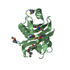
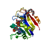
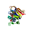
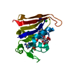
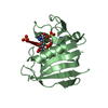
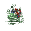
| Entry | Database: PDB / ID: 6xhz | ||||||
|---|---|---|---|---|---|---|---|
| Title | Alpha-lytic protease homolog N4 | ||||||
 Components Components | N4: hypothetical protein | ||||||
 Keywords Keywords | UNKNOWN FUNCTION / Pseudoprotease / trypsin-like fold | ||||||
| Biological species |   Streptomyces monomycini (bacteria) Streptomyces monomycini (bacteria) | ||||||
| Method | X-RAY DIFFRACTION / SYNCHROTRON / MOLECULAR REPLACEMENT / Resolution: 1.25 Å | ||||||
 Authors Authors | Nixon, C.F. / Marqusee, S.M. / Gee, C.L. | ||||||
| Funding support |  United States, 1items United States, 1items
| ||||||
 Citation Citation | Journal: Biochemistry / Year: 2021 Title: Exploring the Evolutionary History of Kinetic Stability in the alpha-Lytic Protease Family. Authors: Nixon, C.F. / Lim, S.A. / Sailer, Z.R. / Zheludev, I.N. / Gee, C.L. / Kelch, B.A. / Harms, M.J. / Marqusee, S. | ||||||
| History |
|
- Structure visualization
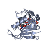
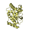
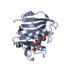
| Structure viewer | Molecule:  MolmilJmol/JSmol MolmilJmol/JSmol |
|---|
- Downloads & links
-Download
| PDBx/mmCIF format | 6xhz.cif.gz | 97.9 KB | Display | PDBx/mmCIF format |
|---|---|---|---|---|
| PDB format | pdb6xhz.ent.gz | 60.4 KB | Display | PDB format |
| PDBx/mmJSON format | 6xhz.json.gz | Tree view | PDBx/mmJSON format | |
| Others |  Other downloads Other downloads |
-Validation report
| Arichive directory | https://data.pdbj.org/pub/pdb/validation_reports/xh/6xhzftp://data.pdbj.org/pub/pdb/validation_reports/xh/6xhz | HTTPS FTP |
|---|
-Related structure data
| Related structure data | 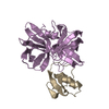 3sgbS S: Starting model for refinement |
|---|---|
| Similar structure data |

-Links
 PDBj
PDBj- Assembly
| Deposited unit | 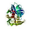
| ||||||||||||
|---|---|---|---|---|---|---|---|---|---|---|---|---|---|
| 1 |
| ||||||||||||
| Unit cell |
|
-Components
| #1: Protein | Mass: 21830.342 Da / Num. of mol.: 1 Source method: isolated from a genetically manipulated source Source: (gene. exp.) Streptomyces monomycini (bacteria) / Gene: hypothetical protein [Streptomyces monomycini]. / Production host: Escherichia coli (E. coli) / Strain (production host): BL21 Rosetta 2(DE3)pLysS |
|---|---|
| #2: Chemical | ChemComp-SO4 / Sulfate  Mass: 96.063 Da / Num. of mol.: 1 / Source method: obtained synthetically / Formula: SO4 Mass: 96.063 Da / Num. of mol.: 1 / Source method: obtained synthetically / Formula: SO4 |
| #3: Water | ChemComp-HOH / Water Mass: 18.015 Da / Num. of mol.: 184 / Source method: isolated from a natural source / Formula: H2O Mass: 18.015 Da / Num. of mol.: 184 / Source method: isolated from a natural source / Formula: H2O |
| Has ligand of interest | N |
-Experimental details
-Experiment
| Experiment | Method: X-RAY DIFFRACTION / Number of used crystals: 1 |
|---|
- Sample preparation
| Crystal | Density Matthews: 1.89 Å3/Da / Density % sol: 34.93 % |
|---|---|
| Crystal grow | Temperature: 293.15 K / Method: vapor diffusion, sitting drop / pH: 7 Details: 1.5 M Lithium sulfate monohydrate, 0.1 M BIS-TRIS propane, pH 7.0 |
-Data collection
| Diffraction | Mean temperature: 100 K / Serial crystal experiment: N |
|---|---|
| Diffraction source | Source: SYNCHROTRON / Site: ALS / Beamline: 8.2.2 / Wavelength: 1 Å |
| Detector | Type: ADSC QUANTUM 315r / Detector: CCD / Date: May 30, 2019 |
| Radiation | Monochromator: S111 / Protocol: SINGLE WAVELENGTH / Monochromatic (M) / Laue (L): M / Scattering type: x-ray |
| Radiation wavelength | Wavelength: 1 Å / Relative weight: 1 |
| Reflection | Resolution: 1.25→41.7 Å / Num. obs: 39417 / % possible obs: 86.6 % / Redundancy: 3.4 % / Biso Wilson estimate: 11.303 Å2 / CC1/2: 0.999 / Rmerge(I) obs: 0.036 / Rpim(I) all: 0.023 / Rrim(I) all: 0.043 / Net I/σ(I): 14.9 |
| Reflection shell | Resolution: 1.25→1.27 Å / Redundancy: 1.5 % / Rmerge(I) obs: 1.034 / Mean I/σ(I) obs: 0.7 / Num. unique obs: 625 / CC1/2: 0.278 / Rpim(I) all: 1.034 / Rrim(I) all: 1.462 / % possible all: 27.6 |
- Processing
| Software |
| |||||||||||||||||||||||||||||||||||||||||||||||||||||||||||||||||||||||||||||||||||||||||||||||||||||||||
|---|---|---|---|---|---|---|---|---|---|---|---|---|---|---|---|---|---|---|---|---|---|---|---|---|---|---|---|---|---|---|---|---|---|---|---|---|---|---|---|---|---|---|---|---|---|---|---|---|---|---|---|---|---|---|---|---|---|---|---|---|---|---|---|---|---|---|---|---|---|---|---|---|---|---|---|---|---|---|---|---|---|---|---|---|---|---|---|---|---|---|---|---|---|---|---|---|---|---|---|---|---|---|---|---|---|---|
| Refinement | Method to determine structure: MOLECULAR REPLACEMENT Starting model: 3SGB Resolution: 1.25→38.68 Å / SU ML: 0.1264 / Cross valid method: FREE R-VALUE / σ(F): 1.34 / Phase error: 23.8137 Stereochemistry target values: GeoStd + Monomer Library + CDL v1.2
| |||||||||||||||||||||||||||||||||||||||||||||||||||||||||||||||||||||||||||||||||||||||||||||||||||||||||
| Solvent computation | Shrinkage radii: 0.9 Å / VDW probe radii: 1.11 Å / Solvent model: FLAT BULK SOLVENT MODEL | |||||||||||||||||||||||||||||||||||||||||||||||||||||||||||||||||||||||||||||||||||||||||||||||||||||||||
| Displacement parameters | Biso mean: 19.93 Å2 | |||||||||||||||||||||||||||||||||||||||||||||||||||||||||||||||||||||||||||||||||||||||||||||||||||||||||
| Refinement step | Cycle: LAST / Resolution: 1.25→38.68 Å
| |||||||||||||||||||||||||||||||||||||||||||||||||||||||||||||||||||||||||||||||||||||||||||||||||||||||||
| Refine LS restraints |
| |||||||||||||||||||||||||||||||||||||||||||||||||||||||||||||||||||||||||||||||||||||||||||||||||||||||||
| LS refinement shell |
|
