 Movie
Movie Controller


+ Open data

- Basic information
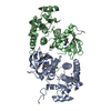
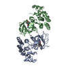
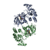
| Entry | Database: PDB / ID: 6xfp | ||||||
|---|---|---|---|---|---|---|---|
| Title | Crystal Structure of BRAF kinase domain bound to Belvarafenib | ||||||
 Components Components | Serine/threonine-protein kinase B-raf | ||||||
 Keywords Keywords |  SIGNALING PROTEIN / BRAF Belvarafenib SIGNALING PROTEIN / BRAF Belvarafenib | ||||||
| Function / homology |  Function and homology informationnon-specific serine/threonine protein kinase / positive regulation of protein phosphorylation / protein phosphorylation / intracellular membrane-bounded organelle / protein serine/threonine kinase activity / negative regulation of apoptotic process / signal transduction / ATP binding / cytosol Function and homology informationnon-specific serine/threonine protein kinase / positive regulation of protein phosphorylation / protein phosphorylation / intracellular membrane-bounded organelle / protein serine/threonine kinase activity / negative regulation of apoptotic process / signal transduction / ATP binding / cytosolSimilarity search - Function | ||||||
| Biological species |  Homo sapiens (human) Homo sapiens (human) | ||||||
| Method | X-RAY DIFFRACTION / SYNCHROTRON / MOLECULAR REPLACEMENT / molecular replacement / Resolution: 2 Å | ||||||
 Authors Authors | Yin, J. / Sudhamsu, J. | ||||||
 Citation Citation | Journal: Nature / Year: 2021 Title: ARAF mutations confer resistance to the RAF inhibitor belvarafenib in melanoma. Authors: Yen, I. / Shanahan, F. / Lee, J. / Hong, Y.S. / Shin, S.J. / Moore, A.R. / Sudhamsu, J. / Chang, M.T. / Bae, I. / Dela Cruz, D. / Hunsaker, T. / Klijn, C. / Liau, N.P.D. / Lin, E. / Martin, ...Authors: Yen, I. / Shanahan, F. / Lee, J. / Hong, Y.S. / Shin, S.J. / Moore, A.R. / Sudhamsu, J. / Chang, M.T. / Bae, I. / Dela Cruz, D. / Hunsaker, T. / Klijn, C. / Liau, N.P.D. / Lin, E. / Martin, S.E. / Modrusan, Z. / Piskol, R. / Segal, E. / Venkatanarayan, A. / Ye, X. / Yin, J. / Zhang, L. / Kim, J.S. / Lim, H.S. / Kim, K.P. / Kim, Y.J. / Han, H.S. / Lee, S.J. / Kim, S.T. / Jung, M. / Hong, Y.H. / Noh, Y.S. / Choi, M. / Han, O. / Nowicka, M. / Srinivasan, S. / Yan, Y. / Kim, T.W. / Malek, S. | ||||||
| History |
|
- Structure visualization
| Structure viewer | Molecule: MolmilJmol/JSmol |
|---|
- Downloads & links
-Download
| PDBx/mmCIF format | 6xfp.cif.gz | 75.3 KB | Display | PDBx/mmCIF format |
|---|---|---|---|---|
| PDB format | pdb6xfp.ent.gz | 52.6 KB | Display | PDB format |
| PDBx/mmJSON format | 6xfp.json.gz | Tree view | PDBx/mmJSON format | |
| Others |  Other downloads Other downloads |
-Validation report
| Arichive directory | https://data.pdbj.org/pub/pdb/validation_reports/xf/6xfpftp://data.pdbj.org/pub/pdb/validation_reports/xf/6xfp | HTTPS FTP |
|---|
-Related structure data
| Related structure data | 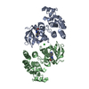 4mnfS S: Starting model for refinement |
|---|---|
| Similar structure data |


-Links
 PDBj
PDBj


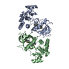
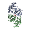
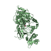
- Assembly
| Deposited unit | 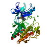
| ||||||||||||
|---|---|---|---|---|---|---|---|---|---|---|---|---|---|
| 1 | 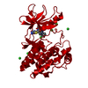
| ||||||||||||
| Unit cell |
| ||||||||||||
| Components on special symmetry positions |
|
-Components
| #1: Protein | Mass: 32832.621 Da / Num. of mol.: 1 Source method: isolated from a genetically manipulated source Source: (gene. exp.) Homo sapiens (human) / Gene: BRAF / Production host:  Escherichia coli (E. coli) / References: UniProt: H7C560 Escherichia coli (E. coli) / References: UniProt: H7C560 | ||||
|---|---|---|---|---|---|
| #2: Chemical | ChemComp-V1Y / Belvarafenib  Mass: 478.929 Da / Num. of mol.: 1 / Source method: obtained synthetically / Formula: C23H16ClFN6OS / Comment: antitumor, inhibitor*YM Mass: 478.929 Da / Num. of mol.: 1 / Source method: obtained synthetically / Formula: C23H16ClFN6OS / Comment: antitumor, inhibitor*YM | ||||
| #3: Chemical | ChemComp-CL / Chloride  Mass: 35.453 Da / Num. of mol.: 6 / Source method: obtained synthetically / Formula: Cl Mass: 35.453 Da / Num. of mol.: 6 / Source method: obtained synthetically / Formula: Cl#4: Water | ChemComp-HOH / | Water Mass: 18.015 Da / Num. of mol.: 86 / Source method: isolated from a natural source / Formula: H2O Mass: 18.015 Da / Num. of mol.: 86 / Source method: isolated from a natural source / Formula: H2OHas ligand of interest | N | |
-Experimental details
-Experiment
| Experiment | Method: X-RAY DIFFRACTION / Number of used crystals: 1 |
|---|
- Sample preparation
| Crystal | Density Matthews: 2.31 Å3/Da / Density % sol: 46.72 % |
|---|---|
| Crystal grow | Temperature: 298 K / Method: vapor diffusion Details: (18% PEG 3350, and 0.2M Na Iodine, and 0.1 M bis-Tris propane pH6.5) |
-Data collection
| Diffraction | Mean temperature: 100 K / Serial crystal experiment: N |
|---|---|
| Diffraction source | Source: SYNCHROTRON / Site: SSRL  / Beamline: BL12-2 / Wavelength: 0.9742 Å / Beamline: BL12-2 / Wavelength: 0.9742 Å |
| Detector | Type: DECTRIS PILATUS 6M / Detector: PIXEL / Date: Jan 22, 2020 |
| Radiation | Protocol: SINGLE WAVELENGTH / Monochromatic (M) / Laue (L): M / Scattering type: x-ray |
| Radiation wavelength | Wavelength: 0.9742 Å / Relative weight: 1 |
| Reflection | Resolution: 2→59.782 Å / Num. obs: 20506 / % possible obs: 95.62 % / Redundancy: 5.9 % / Biso Wilson estimate: 32.24 Å2 / CC1/2: 0.999 / Net I/σ(I): 12.64 |
| Reflection shell | Resolution: 2→2.07 Å / Num. unique obs: 1961 / CC1/2: 0.636 |
-Phasing
| Phasing | Method: molecular replacement |
|---|
- Processing
| Software |
| ||||||||||||||||||||||||||||||||||||||||||||||||||||||||||||
|---|---|---|---|---|---|---|---|---|---|---|---|---|---|---|---|---|---|---|---|---|---|---|---|---|---|---|---|---|---|---|---|---|---|---|---|---|---|---|---|---|---|---|---|---|---|---|---|---|---|---|---|---|---|---|---|---|---|---|---|---|---|
| Refinement | Method to determine structure: MOLECULAR REPLACEMENT Starting model: 4MNF Resolution: 2→59.782 Å / SU ML: 0.27 / Cross valid method: THROUGHOUT / σ(F): 1.33 / Phase error: 29.68 / Stereochemistry target values: ML
| ||||||||||||||||||||||||||||||||||||||||||||||||||||||||||||
| Solvent computation | Shrinkage radii: 0.9 Å / VDW probe radii: 1.11 Å / Solvent model: FLAT BULK SOLVENT MODEL | ||||||||||||||||||||||||||||||||||||||||||||||||||||||||||||
| Displacement parameters | Biso max: 93.84 Å2 / Biso mean: 39.9297 Å2 / Biso min: 13.02 Å2 | ||||||||||||||||||||||||||||||||||||||||||||||||||||||||||||
| Refinement step | Cycle: final / Resolution: 2→59.782 Å
| ||||||||||||||||||||||||||||||||||||||||||||||||||||||||||||
| Refine LS restraints |
| ||||||||||||||||||||||||||||||||||||||||||||||||||||||||||||
| LS refinement shell | Refine-ID: X-RAY DIFFRACTION / Rfactor Rfree error: 0
|
