 Movie
Movie Controller

[English] 日本語
 Yorodumi
- PDB-6xfi: Crystal Structures of beta-1,4-N-Acetylglucosaminyltransferase 2 ... -
+ Open data

- Basic information
| Entry | Database: PDB / ID: 6xfi | ||||||
|---|---|---|---|---|---|---|---|
| Title | Crystal Structures of beta-1,4-N-Acetylglucosaminyltransferase 2 (POMGNT2): Structural Basis for Inherited Muscular Dystrophies | ||||||
 Components Components | Protein O-linked-mannose beta-1,4-N-acetylglucosaminyltransferase 2 | ||||||
 Keywords Keywords |  TRANSFERASE / muscular dystrophy / alpha-dystroglycan / O-mannosylation / POMGNT2 TRANSFERASE / muscular dystrophy / alpha-dystroglycan / O-mannosylation / POMGNT2 | ||||||
| Function / homology |  Function and homology information Function and homology informationprotein O-mannose beta-1,4-N-acetylglucosaminyltransferase / O-linked glycosylation / protein O-linked mannosylation / protein O-acetylglucosaminyltransferase activity / acetylglucosaminyltransferase activity / protein O-linked glycosylation / glycosyltransferase activity / neuron migration / endoplasmic reticulum membrane / endoplasmic reticulumSimilarity search - Function | ||||||
| Biological species |  Homo sapiens (human) Homo sapiens (human) | ||||||
| Method | X-RAY DIFFRACTION / SYNCHROTRON / SAD / Resolution: 2 Å | ||||||
 Authors Authors | Halmo, S.M. / Yeh, J. / Wells, L. / Moremen, K.W. / Lanzilotta, W.N. | ||||||
| Funding support |  United States, 1items United States, 1items
| ||||||
 Citation Citation | Journal: Acta Crystallogr D Struct Biol / Year: 2021 Title: Crystal structures of beta-1,4-N-acetylglucosaminyltransferase 2: structural basis for inherited muscular dystrophies. Authors: Yang, J.Y. / Halmo, S.M. / Praissman, J. / Chapla, D. / Singh, D. / Wells, L. / Moremen, K.W. / Lanzilotta, W.N. | ||||||
| History |
|
- Structure visualization
| Structure viewer | Molecule: MolmilJmol/JSmol |
|---|
- Downloads & links
-Download
| PDBx/mmCIF format | 6xfi.cif.gz | 122.6 KB | Display | PDBx/mmCIF format |
|---|---|---|---|---|
| PDB format | pdb6xfi.ent.gz | 97.6 KB | Display | PDB format |
| PDBx/mmJSON format | 6xfi.json.gz | Tree view | PDBx/mmJSON format | |
| Others |  Other downloads Other downloads |
-Validation report
| Arichive directory | https://data.pdbj.org/pub/pdb/validation_reports/xf/6xfiftp://data.pdbj.org/pub/pdb/validation_reports/xf/6xfi | HTTPS FTP |
|---|
-Related structure data


-Links
 PDBj
PDBj


- Assembly
| Deposited unit | 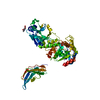
| ||||||||
|---|---|---|---|---|---|---|---|---|---|
| 1 | 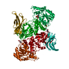
| ||||||||
| Unit cell |
|
-Components
| #1: Protein | Mass: 61062.449 Da / Num. of mol.: 1 / Fragment: UNP residues 52-580 Source method: isolated from a genetically manipulated source Source: (gene. exp.) Homo sapiens (human) / Gene: POMGNT2, AGO61, C3orf39, EOGTL, GTDC2 / Cell line (production host): HEK293S / Production host: Homo sapiens (human)References: UniProt: Q8NAT1, protein O-mannose beta-1,4-N-acetylglucosaminyltransferase | ||||
|---|---|---|---|---|---|
| #2: Chemical | ChemComp-UDP / Uridine diphosphate  Type: RNA linking / Mass: 404.161 Da / Num. of mol.: 1 / Source method: obtained synthetically / Formula: C9H14N2O12P2 / Comment: UDP*YM Type: RNA linking / Mass: 404.161 Da / Num. of mol.: 1 / Source method: obtained synthetically / Formula: C9H14N2O12P2 / Comment: UDP*YM | ||||
| #3: Sugar | ChemComp-NAG / N-Acetylglucosamine  Type: D-saccharide, beta linking / Mass: 221.208 Da / Num. of mol.: 1 Type: D-saccharide, beta linking / Mass: 221.208 Da / Num. of mol.: 1Source method: isolated from a genetically manipulated source Formula: C8H15NO6 | ||||
| #4: Chemical | Phosphate  Mass: 94.971 Da / Num. of mol.: 2 / Source method: obtained synthetically / Formula: PO4 Mass: 94.971 Da / Num. of mol.: 2 / Source method: obtained synthetically / Formula: PO4#5: Water | ChemComp-HOH / | Water Mass: 18.015 Da / Num. of mol.: 194 / Source method: isolated from a natural source / Formula: H2O Mass: 18.015 Da / Num. of mol.: 194 / Source method: isolated from a natural source / Formula: H2OHas ligand of interest | N | |
-Experimental details
-Experiment
| Experiment | Method: X-RAY DIFFRACTION / Number of used crystals: 1 |
|---|
- Sample preparation
| Crystal | Density Matthews: 2.81 Å3/Da / Density % sol: 56.3 % |
|---|---|
| Crystal grow | Temperature: 296 K / Method: vapor diffusion, hanging drop Details: 0.1 M potassium/sodium tartrate, 0.1 M Bis-Tris, pH 7.5, 10% PEG10000 |
-Data collection
| Diffraction | Mean temperature: 100 K / Serial crystal experiment: N |
|---|---|
| Diffraction source | Source: SYNCHROTRON / Site: APS / Beamline: 22-ID / Wavelength: 1 Å |
| Detector | Type: DECTRIS EIGER X 16M / Detector: PIXEL / Date: Apr 6, 2019 |
| Radiation | Monochromator: double crystal Si(111) / Protocol: SINGLE WAVELENGTH / Monochromatic (M) / Laue (L): M / Scattering type: x-ray |
| Radiation wavelength | Wavelength: 1 Å / Relative weight: 1 |
| Reflection | Resolution: 1.97→50 Å / Num. obs: 49420 / % possible obs: 99.7 % / Redundancy: 11.3 % / CC1/2: 1 / CC star: 1 / Net I/σ(I): 27.5 |
| Reflection shell | Resolution: 1.97→2.08 Å / Num. unique obs: 4759 / CC1/2: 0.298 / CC star: 0.678 |
- Processing
| Software |
| |||||||||||||||||||||||||||||||||||||||||||||||||||||||||||||||||||||||||||||||||||||||||||||||||||||||||
|---|---|---|---|---|---|---|---|---|---|---|---|---|---|---|---|---|---|---|---|---|---|---|---|---|---|---|---|---|---|---|---|---|---|---|---|---|---|---|---|---|---|---|---|---|---|---|---|---|---|---|---|---|---|---|---|---|---|---|---|---|---|---|---|---|---|---|---|---|---|---|---|---|---|---|---|---|---|---|---|---|---|---|---|---|---|---|---|---|---|---|---|---|---|---|---|---|---|---|---|---|---|---|---|---|---|---|
| Refinement | Method to determine structure: SAD / Resolution: 2→40 Å / SU ML: 0.26 / Cross valid method: THROUGHOUT / σ(F): 1.33 / Phase error: 23.74 / Stereochemistry target values: ML
| |||||||||||||||||||||||||||||||||||||||||||||||||||||||||||||||||||||||||||||||||||||||||||||||||||||||||
| Solvent computation | Shrinkage radii: 0.9 Å / VDW probe radii: 1.11 Å / Solvent model: FLAT BULK SOLVENT MODEL | |||||||||||||||||||||||||||||||||||||||||||||||||||||||||||||||||||||||||||||||||||||||||||||||||||||||||
| Displacement parameters | Biso max: 107.17 Å2 / Biso mean: 40.1046 Å2 / Biso min: 16.41 Å2 | |||||||||||||||||||||||||||||||||||||||||||||||||||||||||||||||||||||||||||||||||||||||||||||||||||||||||
| Refinement step | Cycle: final / Resolution: 2→40 Å
| |||||||||||||||||||||||||||||||||||||||||||||||||||||||||||||||||||||||||||||||||||||||||||||||||||||||||
| LS refinement shell | Refine-ID: X-RAY DIFFRACTION / Rfactor Rfree error: 0 / Total num. of bins used: 14
|
