 Movie
Movie Controller


+ Open data

- Basic information
| Entry | Database: PDB / ID: 7e9j | |||||||||||||||
|---|---|---|---|---|---|---|---|---|---|---|---|---|---|---|---|---|
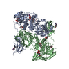
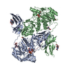
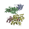
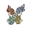
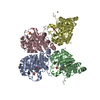
| Title | Crystal Structure of POMGNT2 in complex with UDP | |||||||||||||||
 Components Components | Protein O-linked-mannose beta-1,4-N-acetylglucosaminyltransferase 2 | |||||||||||||||
 Keywords Keywords |  TRANSFERASE / Glycosyltransferase / O-mannose type glycosylation TRANSFERASE / Glycosyltransferase / O-mannose type glycosylation | |||||||||||||||
| Function / homology |  Function and homology informationO-linked glycosylation / protein O-mannose beta-1,4-N-acetylglucosaminyltransferase / protein O-linked mannosylation / protein O-acetylglucosaminyltransferase activity / acetylglucosaminyltransferase activity / protein O-linked glycosylation / neuron migration / endoplasmic reticulum membrane / endoplasmic reticulum Function and homology informationO-linked glycosylation / protein O-mannose beta-1,4-N-acetylglucosaminyltransferase / protein O-linked mannosylation / protein O-acetylglucosaminyltransferase activity / acetylglucosaminyltransferase activity / protein O-linked glycosylation / neuron migration / endoplasmic reticulum membrane / endoplasmic reticulumSimilarity search - Function | |||||||||||||||
| Biological species |  Bos taurus (cattle) Bos taurus (cattle) | |||||||||||||||
| Method | X-RAY DIFFRACTION / SYNCHROTRON / SAD / Resolution: 2.4 Å | |||||||||||||||
 Authors Authors | Kuwabara, N. | |||||||||||||||
| Funding support |  Japan, 4items Japan, 4items
| |||||||||||||||
 Citation Citation | Journal: Genes Cells / Year: 2021 Title: The structure of POMGNT2 provides new insights into the mechanism to determine the functional O-mannosylation site on alpha-dystroglycan. Authors: Imae, R. / Kuwabara, N. / Manya, H. / Tanaka, T. / Tsuyuguchi, M. / Mizuno, M. / Endo, T. / Kato, R. | |||||||||||||||
| History |
|
- Structure visualization
| Structure viewer | Molecule: MolmilJmol/JSmol |
|---|
- Downloads & links
-Download
| PDBx/mmCIF format | 7e9j.cif.gz | 514.7 KB | Display | PDBx/mmCIF format |
|---|---|---|---|---|
| PDB format | pdb7e9j.ent.gz | 364.7 KB | Display | PDB format |
| PDBx/mmJSON format | 7e9j.json.gz | Tree view | PDBx/mmJSON format | |
| Others |  Other downloads Other downloads |
-Validation report
| Arichive directory | https://data.pdbj.org/pub/pdb/validation_reports/e9/7e9jftp://data.pdbj.org/pub/pdb/validation_reports/e9/7e9j | HTTPS FTP |
|---|
-Related structure data


-Links
 PDBj
PDBj


- Assembly
| Deposited unit | 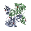
| ||||||||||||
|---|---|---|---|---|---|---|---|---|---|---|---|---|---|
| 1 |
| ||||||||||||
| Unit cell |
|
-Components
| #1: Protein | Mass: 61563.902 Da / Num. of mol.: 2 Source method: isolated from a genetically manipulated source Source: (gene. exp.) Bos taurus (cattle) / Gene: POMGNT2, AGO61, GTDC2 / Production host:  Homo sapiens (human) Homo sapiens (human)References: UniProt: Q5NDF2, protein O-mannose beta-1,4-N-acetylglucosaminyltransferase #2: Sugar | ChemComp-NAG / N-Acetylglucosamine  Type: D-saccharide, beta linking / Mass: 221.208 Da / Num. of mol.: 4 / Source method: obtained synthetically / Formula: C8H15NO6 Type: D-saccharide, beta linking / Mass: 221.208 Da / Num. of mol.: 4 / Source method: obtained synthetically / Formula: C8H15NO6#3: Chemical | Uridine diphosphate  Type: RNA linking / Mass: 404.161 Da / Num. of mol.: 2 / Source method: obtained synthetically / Formula: C9H14N2O12P2 / Feature type: SUBJECT OF INVESTIGATION / Comment: UDP*YM Type: RNA linking / Mass: 404.161 Da / Num. of mol.: 2 / Source method: obtained synthetically / Formula: C9H14N2O12P2 / Feature type: SUBJECT OF INVESTIGATION / Comment: UDP*YM#4: Chemical | ChemComp-TRS / | Tris  Mass: 122.143 Da / Num. of mol.: 1 / Source method: obtained synthetically / Formula: C4H12NO3 / Feature type: SUBJECT OF INVESTIGATION / Comment: pH buffer*YM Mass: 122.143 Da / Num. of mol.: 1 / Source method: obtained synthetically / Formula: C4H12NO3 / Feature type: SUBJECT OF INVESTIGATION / Comment: pH buffer*YM#5: Water | ChemComp-HOH / | Water Mass: 18.015 Da / Num. of mol.: 242 / Source method: isolated from a natural source / Formula: H2O Mass: 18.015 Da / Num. of mol.: 242 / Source method: isolated from a natural source / Formula: H2OHas ligand of interest | Y | |
|---|
-Experimental details
-Experiment
| Experiment | Method: X-RAY DIFFRACTION / Number of used crystals: 1 |
|---|
- Sample preparation
| Crystal | Density Matthews: 2.74 Å3/Da / Density % sol: 55.18 % |
|---|---|
| Crystal grow | Temperature: 277 K / Method: vapor diffusion, hanging drop Details: 0.1 M Tris-HCl (pH 8.0 or 8.5), 12-16 % PEG 8000, 1 % tacsimate (pH 7.0) PH range: 8.0-8.5 |
-Data collection
| Diffraction | Mean temperature: 100 K / Serial crystal experiment: N |
|---|---|
| Diffraction source | Source: SYNCHROTRON / Site: Photon Factory / Beamline: BL-17A / Wavelength: 1.0718 Å |
| Detector | Type: DECTRIS PILATUS 6M / Detector: PIXEL / Date: Nov 17, 2016 |
| Radiation | Protocol: SINGLE WAVELENGTH / Monochromatic (M) / Laue (L): M / Scattering type: x-ray |
| Radiation wavelength | Wavelength: 1.0718 Å / Relative weight: 1 |
| Reflection | Resolution: 2.4→49.3 Å / Num. obs: 56436 / % possible obs: 100 % / Redundancy: 13.4 % / Biso Wilson estimate: 42.4 Å2 / CC1/2: 0.997 / Rmerge(I) obs: 0.247 / Rpim(I) all: 0.07 / Net I/σ(I): 10.5 |
| Reflection shell | Resolution: 2.4→2.486 Å / Rmerge(I) obs: 1.858 / Num. unique obs: 5292 / CC1/2: 0.442 / Rpim(I) all: 0.518 |
- Processing
| Software |
| |||||||||||||||||||||||||||||||||||||||||||||||||||||||||||||||||||||||||||||||||||||||||||||||||||||||||||||||||||||||||||||||||||||||||||||||||||||||||||||||||||||||||||||||
|---|---|---|---|---|---|---|---|---|---|---|---|---|---|---|---|---|---|---|---|---|---|---|---|---|---|---|---|---|---|---|---|---|---|---|---|---|---|---|---|---|---|---|---|---|---|---|---|---|---|---|---|---|---|---|---|---|---|---|---|---|---|---|---|---|---|---|---|---|---|---|---|---|---|---|---|---|---|---|---|---|---|---|---|---|---|---|---|---|---|---|---|---|---|---|---|---|---|---|---|---|---|---|---|---|---|---|---|---|---|---|---|---|---|---|---|---|---|---|---|---|---|---|---|---|---|---|---|---|---|---|---|---|---|---|---|---|---|---|---|---|---|---|---|---|---|---|---|---|---|---|---|---|---|---|---|---|---|---|---|---|---|---|---|---|---|---|---|---|---|---|---|---|---|---|---|---|
| Refinement | Method to determine structure: SAD / Resolution: 2.4→49.3 Å / SU ML: 0.321 / Cross valid method: FREE R-VALUE / σ(F): 1.34 / Phase error: 26.0105 Stereochemistry target values: GeoStd + Monomer Library + CDL v1.2
| |||||||||||||||||||||||||||||||||||||||||||||||||||||||||||||||||||||||||||||||||||||||||||||||||||||||||||||||||||||||||||||||||||||||||||||||||||||||||||||||||||||||||||||||
| Solvent computation | Shrinkage radii: 0.9 Å / VDW probe radii: 1.11 Å / Solvent model: FLAT BULK SOLVENT MODEL | |||||||||||||||||||||||||||||||||||||||||||||||||||||||||||||||||||||||||||||||||||||||||||||||||||||||||||||||||||||||||||||||||||||||||||||||||||||||||||||||||||||||||||||||
| Displacement parameters | Biso mean: 54.85 Å2 | |||||||||||||||||||||||||||||||||||||||||||||||||||||||||||||||||||||||||||||||||||||||||||||||||||||||||||||||||||||||||||||||||||||||||||||||||||||||||||||||||||||||||||||||
| Refinement step | Cycle: LAST / Resolution: 2.4→49.3 Å
| |||||||||||||||||||||||||||||||||||||||||||||||||||||||||||||||||||||||||||||||||||||||||||||||||||||||||||||||||||||||||||||||||||||||||||||||||||||||||||||||||||||||||||||||
| Refine LS restraints |
| |||||||||||||||||||||||||||||||||||||||||||||||||||||||||||||||||||||||||||||||||||||||||||||||||||||||||||||||||||||||||||||||||||||||||||||||||||||||||||||||||||||||||||||||
| LS refinement shell |
| |||||||||||||||||||||||||||||||||||||||||||||||||||||||||||||||||||||||||||||||||||||||||||||||||||||||||||||||||||||||||||||||||||||||||||||||||||||||||||||||||||||||||||||||
| Refinement TLS params. | Method: refined / Refine-ID: X-RAY DIFFRACTION
| |||||||||||||||||||||||||||||||||||||||||||||||||||||||||||||||||||||||||||||||||||||||||||||||||||||||||||||||||||||||||||||||||||||||||||||||||||||||||||||||||||||||||||||||
| Refinement TLS group |
|
