Movie
Movie Controller

[English] 日本語
 Yorodumi
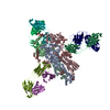
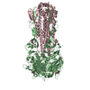
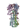
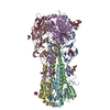
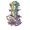
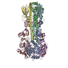
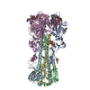
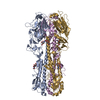
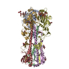
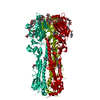
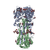
- PDB-6wxb: Cryo-EM Structure of Influenza Hemagglutinin (HA) Trimer Vitrifie... -
+ Open data

- Basic information
| Entry | Database: PDB / ID: 6wxb | ||||||||||||
|---|---|---|---|---|---|---|---|---|---|---|---|---|---|
| Title | Cryo-EM Structure of Influenza Hemagglutinin (HA) Trimer Vitrified Using Back-it-up | ||||||||||||
 Components Components | Hemagglutinin | ||||||||||||
 Keywords Keywords | VIRAL PROTEIN / Hemagglutinin / trimer / Influenza / back-it-up / through-grid wicking | ||||||||||||
| Function / homology |  Function and homology informationviral budding from plasma membrane / clathrin-dependent endocytosis of virus by host cell / host cell surface receptor binding / fusion of virus membrane with host plasma membrane / fusion of virus membrane with host endosome membrane / viral envelope / virion attachment to host cell / host cell plasma membrane / virion membrane / membrane Function and homology informationviral budding from plasma membrane / clathrin-dependent endocytosis of virus by host cell / host cell surface receptor binding / fusion of virus membrane with host plasma membrane / fusion of virus membrane with host endosome membrane / viral envelope / virion attachment to host cell / host cell plasma membrane / virion membrane / membraneSimilarity search - Function | ||||||||||||
| Biological species |   Influenza A virus Influenza A virus | ||||||||||||
| Method | ELECTRON MICROSCOPY / single particle reconstruction / cryo EM / Resolution: 2.9 Å | ||||||||||||
 Authors Authors | Tan, Y.Z. / Rubinstein, J.L. | ||||||||||||
| Funding support |  Canada, 3items Canada, 3items
| ||||||||||||
 Citation Citation | Journal: Acta Crystallogr D Struct Biol / Year: 2020 Title: Through-grid wicking enables high-speed cryoEM specimen preparation. Authors: Yong Zi Tan / John L Rubinstein / Abstract: Blotting times for conventional cryoEM specimen preparation complicate time-resolved studies and lead to some specimens adopting preferred orientations or denaturing at the air-water interface. Here, ...Blotting times for conventional cryoEM specimen preparation complicate time-resolved studies and lead to some specimens adopting preferred orientations or denaturing at the air-water interface. Here, it is shown that solution sprayed onto one side of a holey cryoEM grid can be wicked through the grid by a glass-fiber filter held against the opposite side, often called the `back', of the grid, producing a film suitable for vitrification. This process can be completed in tens of milliseconds. Ultrasonic specimen application and through-grid wicking were combined in a high-speed specimen-preparation device that was named `Back-it-up' or BIU. The high liquid-absorption capacity of the glass fiber compared with self-wicking grids makes the method relatively insensitive to the amount of sample applied. Consequently, through-grid wicking produces large areas of ice that are suitable for cryoEM for both soluble and detergent-solubilized protein complexes. The speed of the device increases the number of views for a specimen that suffers from preferred orientations. | ||||||||||||
| History |
|
- Structure visualization
| Movie |
Movie viewer |
|---|---|
| Structure viewer | Molecule: MolmilJmol/JSmol |
- Downloads & links
-Download
| PDBx/mmCIF format | 6wxb.cif.gz | 471.3 KB | Display | PDBx/mmCIF format |
|---|---|---|---|---|
| PDB format | pdb6wxb.ent.gz | 408.4 KB | Display | PDB format |
| PDBx/mmJSON format | 6wxb.json.gz | Tree view | PDBx/mmJSON format | |
| Others |  Other downloads Other downloads |
-Validation report
| Arichive directory | https://data.pdbj.org/pub/pdb/validation_reports/wx/6wxbftp://data.pdbj.org/pub/pdb/validation_reports/wx/6wxb | HTTPS FTP |
|---|
-Related structure data
| Related structure data | 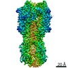 21954MC  6wx6C M: map data used to model this data C: citing same article ( |
|---|---|
| Similar structure data | |
| EM raw data | EMPIAR-10532 (Title: Single-Particle CryoEM of Influenza Hemagglutinin (HA) Trimer Vitrified Using Back-it-up Data size: 1.2 TB Data #1: Unaligned Falcon IV movies [micrographs - multiframe] Data #2: Aligned micrographs [micrographs - single frame] Data #3: Final Refined Particles with Euler Angles and Shifts [picked particles - single frame - processed]) |


-Links
 PDBj
PDBj


- Assembly
| Deposited unit | 
|
|---|---|
| 1 |
|
-Components
| #1: Protein | Mass: 62855.215 Da / Num. of mol.: 3 Source method: isolated from a genetically manipulated source Source: (gene. exp.) Influenza A virus (strain A/Hong Kong/1/1968 H3N2)Strain: A/Hong Kong/1/1968 H3N2 / Gene: HA / Production host:  Homo sapiens (human) / Strain (production host): HEK293S / References: UniProt: Q91MA7 Homo sapiens (human) / Strain (production host): HEK293S / References: UniProt: Q91MA7#2: Sugar | ChemComp-NAG / N-Acetylglucosamine  Type: D-saccharide, beta linking / Mass: 221.208 Da / Num. of mol.: 12 Type: D-saccharide, beta linking / Mass: 221.208 Da / Num. of mol.: 12Source method: isolated from a genetically manipulated source Formula: C8H15NO6 #3: Sugar | Mannose  Type: D-saccharide, beta linking / Mass: 180.156 Da / Num. of mol.: 3 Type: D-saccharide, beta linking / Mass: 180.156 Da / Num. of mol.: 3Source method: isolated from a genetically manipulated source Formula: C6H12O6 Has ligand of interest | N | |
|---|
-Experimental details
-Experiment
| Experiment | Method: ELECTRON MICROSCOPY |
|---|---|
| EM experiment | Aggregation state: PARTICLE / 3D reconstruction method: single particle reconstruction |
- Sample preparation
| Component | Name: Influenza Hemagglutinin Trimer (H3N2) (A/Hong Kong/1/1968) Type: ORGANELLE OR CELLULAR COMPONENT / Entity ID: #1 / Source: RECOMBINANT |
|---|---|
| Molecular weight | Value: 0.19 MDa / Experimental value: NO |
| Source (natural) | Organism: H3N2 subtype (virus) |
| Source (recombinant) | Organism: Homo sapiens (human) / Strain: HEK293F |
| Buffer solution | pH: 7.4 |
| Specimen | Embedding applied: NO / Shadowing applied: NO / Staining applied: NO / Vitrification applied: YES |
| Specimen support | Details: Grid was glow discharged on both sides for 120s each. Grid material: COPPER/RHODIUM / Grid type: Homemade |
| Vitrification | Instrument: HOMEMADE PLUNGER / Cryogen name: ETHANE-PROPANE / Humidity: 50 % / Chamber temperature: 298 K Details: Back-it-up (ultrasonic specimen application and through-grid wicking in a high-speed specimen preparation device) was used |
- Electron microscopy imaging
| Experimental equipment |  Model: Titan Krios / Image courtesy: FEI Company |
|---|---|
| Microscopy | Model: FEI TITAN KRIOS |
| Electron gun | Electron source: FIELD EMISSION GUN / Accelerating voltage: 300 kV / Illumination mode: FLOOD BEAM |
| Electron lens | Mode: BRIGHT FIELDBright-field microscopy / Cs: 2.7 mm / Alignment procedure: ZEMLIN TABLEAU |
| Specimen holder | Cryogen: NITROGEN / Specimen holder model: FEI TITAN KRIOS AUTOGRID HOLDER |
| Image recording | Average exposure time: 9 sec. / Electron dose: 45 e/Å2 / Film or detector model: FEI FALCON IV (4k x 4k) / Num. of grids imaged: 1 / Num. of real images: 1556 |
| Image scans | Sampling size: 14 µm / Width: 4096 / Height: 4096 |
- Processing
| Software |
| ||||||||||||||||||||||||||||||||||||||||
|---|---|---|---|---|---|---|---|---|---|---|---|---|---|---|---|---|---|---|---|---|---|---|---|---|---|---|---|---|---|---|---|---|---|---|---|---|---|---|---|---|---|
| EM software |
| ||||||||||||||||||||||||||||||||||||||||
| CTF correction | Type: PHASE FLIPPING AND AMPLITUDE CORRECTION | ||||||||||||||||||||||||||||||||||||||||
| Particle selection | Num. of particles selected: 378143 | ||||||||||||||||||||||||||||||||||||||||
| Symmetry | Point symmetry: C3 (3 fold cyclic) | ||||||||||||||||||||||||||||||||||||||||
| 3D reconstruction | Resolution: 2.9 Å / Resolution method: FSC 0.143 CUT-OFF / Num. of particles: 128305 / Symmetry type: POINT | ||||||||||||||||||||||||||||||||||||||||
| Atomic model building | Protocol: RIGID BODY FIT / Space: REAL | ||||||||||||||||||||||||||||||||||||||||
| Atomic model building | PDB-ID: 3WHE | ||||||||||||||||||||||||||||||||||||||||
| Refinement | Cross valid method: NONE / Stereochemistry target values: CDL v1.2 | ||||||||||||||||||||||||||||||||||||||||
| Refine LS restraints |
|