 Movie
Movie Controller


+ Open data

- Basic information
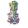
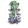
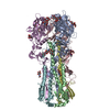
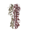
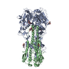
| Entry | Database: PDB / ID: 1mql | |||||||||
|---|---|---|---|---|---|---|---|---|---|---|
| Title | BHA of Ukr/63 | |||||||||
 Components Components | (Hemagglutinin ... ) x 2 ) x 2 | |||||||||
 Keywords Keywords | VIRAL PROTEIN / influenza a virus | |||||||||
| Function / homology |  Function and homology informationviral budding from plasma membrane / clathrin-dependent endocytosis of virus by host cell / host cell surface receptor binding / fusion of virus membrane with host plasma membrane / fusion of virus membrane with host endosome membrane / viral envelope / virion attachment to host cell / host cell plasma membrane / virion membrane / membrane Function and homology informationviral budding from plasma membrane / clathrin-dependent endocytosis of virus by host cell / host cell surface receptor binding / fusion of virus membrane with host plasma membrane / fusion of virus membrane with host endosome membrane / viral envelope / virion attachment to host cell / host cell plasma membrane / virion membrane / membraneSimilarity search - Function | |||||||||
| Biological species |   Influenza A virus Influenza A virus | |||||||||
| Method | X-RAY DIFFRACTION / SYNCHROTRON / MOLECULAR REPLACEMENT / Resolution: 2.9 Å | |||||||||
 Authors Authors | ha, y. / stevens, d.j. / skehel, j.j. / wiley, d.c. | |||||||||
 Citation Citation | Journal: Virology / Year: 2003 Title: X-ray structure of the hemagglutinin of a potential H3 avian progenitor of the 1968 Hong Kong pandemic influenza virus. Authors: Ha, Y. / Stevens, D.J. / Skehel, J.J. / Wiley, D.C. | |||||||||
| History |
|
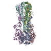
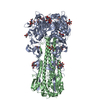
- Structure visualization
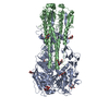
| Structure viewer | Molecule: MolmilJmol/JSmol |
|---|
- Downloads & links
-Download
| PDBx/mmCIF format | 1mql.cif.gz | 293.8 KB | Display | PDBx/mmCIF format |
|---|---|---|---|---|
| PDB format | pdb1mql.ent.gz | 245.1 KB | Display | PDB format |
| PDBx/mmJSON format | 1mql.json.gz | Tree view | PDBx/mmJSON format | |
| Others |  Other downloads Other downloads |
-Validation report
| Arichive directory | https://data.pdbj.org/pub/pdb/validation_reports/mq/1mqlftp://data.pdbj.org/pub/pdb/validation_reports/mq/1mql | HTTPS FTP |
|---|
-Related structure data


-Links
 PDBj
PDBj


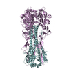
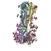
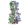
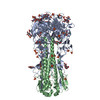
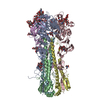
- Assembly
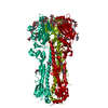
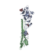
| Deposited unit | 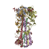
| ||||||||
|---|---|---|---|---|---|---|---|---|---|
| 1 |
| ||||||||
| 2 | 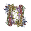
| ||||||||
| Unit cell |
|
-Components
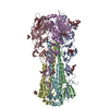
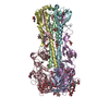
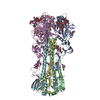
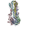
-Hemagglutinin ... , 2 types, 6 molecules ADGBEH
| #1: Protein | Mass: 36285.672 Da / Num. of mol.: 3 / Fragment: residues 17-345 / Source method: isolated from a natural source / Source: (natural) Influenza A virus / Genus: Influenzavirus A / References: UniProt: P03442#2: Protein | Mass: 25574.973 Da / Num. of mol.: 3 / Fragment: residues 346-566 / Source method: isolated from a natural source / Source: (natural) Influenza A virus / Genus: Influenzavirus A / References: UniProt: P03442 |
|---|
-Sugars , 3 types, 25 molecules 


| #3: Sugar | ChemComp-NAG / N-Acetylglucosamine Type: D-saccharide, beta linking / Mass: 221.208 Da / Num. of mol.: 7 Type: D-saccharide, beta linking / Mass: 221.208 Da / Num. of mol.: 7Source method: isolated from a genetically manipulated source Formula: C8H15NO6 #4: Sugar | ChemComp-NDG / N-Acetylglucosamine Type: D-saccharide, alpha linking / Mass: 221.208 Da / Num. of mol.: 12 Type: D-saccharide, alpha linking / Mass: 221.208 Da / Num. of mol.: 12Source method: isolated from a genetically manipulated source Formula: C8H15NO6 #5: Sugar | ChemComp-MAN / Mannose Type: D-saccharide, alpha linking / Mass: 180.156 Da / Num. of mol.: 6 Type: D-saccharide, alpha linking / Mass: 180.156 Da / Num. of mol.: 6Source method: isolated from a genetically manipulated source Formula: C6H12O6 |
|---|
-Non-polymers , 1 types, 47 molecules 
| #6: Water | ChemComp-HOH / WaterMass: 18.015 Da / Num. of mol.: 47 / Source method: isolated from a natural source / Formula: H2O |
|---|
-Experimental details
-Experiment
| Experiment | Method: X-RAY DIFFRACTION / Number of used crystals: 1 |
|---|
- Sample preparation
| Crystal | Density Matthews: 3.69 Å3/Da / Density % sol: 66.63 % | ||||||||||||||||||||||||||||||
|---|---|---|---|---|---|---|---|---|---|---|---|---|---|---|---|---|---|---|---|---|---|---|---|---|---|---|---|---|---|---|---|
| Crystal grow | Temperature: 298 K / Method: vapor diffusion, hanging drop / pH: 7.5 Details: Na citrate, pH 7.5, VAPOR DIFFUSION, HANGING DROP, temperature 298.0K | ||||||||||||||||||||||||||||||
| Crystal grow | *PLUS Method: vapor diffusion, hanging drop | ||||||||||||||||||||||||||||||
| Components of the solutions | *PLUS
|
-Data collection
| Diffraction | Mean temperature: 100 K |
|---|---|
| Diffraction source | Source: SYNCHROTRON / Site: APS  / Beamline: 14-BM-C / Wavelength: 1 Å / Beamline: 14-BM-C / Wavelength: 1 Å |
| Detector | Type: ADSC QUANTUM 4 / Detector: CCD / Date: Jan 1, 2000 / Details: mirrors |
| Radiation | Monochromator: graphite / Protocol: SINGLE WAVELENGTH / Monochromatic (M) / Laue (L): M / Scattering type: x-ray |
| Radiation wavelength | Wavelength: 1 Å / Relative weight: 1 |
| Reflection | Resolution: 2.9→40 Å / Num. all: 59854 / Num. obs: 58957 / % possible obs: 98.5 % / Observed criterion σ(F): 2 / Observed criterion σ(I): 2 / Redundancy: 4 % / Biso Wilson estimate: 50 Å2 / Rmerge(I) obs: 0.1 / Rsym value: 0.1 / Net I/σ(I): 8 |
| Reflection shell | Highest resolution: 2.9 Å / % possible all: 98.5 |
| Reflection | *PLUS Lowest resolution: 40 Å / Redundancy: 4.3 % / Rmerge(I) obs: 0.1 |
| Reflection shell | *PLUS % possible obs: 96 % / Rmerge(I) obs: 0.549 |
- Processing
| Software |
| ||||||||||||||||||||
|---|---|---|---|---|---|---|---|---|---|---|---|---|---|---|---|---|---|---|---|---|---|
| Refinement | Method to determine structure: MOLECULAR REPLACEMENT / Resolution: 2.9→40 Å / σ(F): 0 / Stereochemistry target values: Engh & Huber
| ||||||||||||||||||||
| Refinement step | Cycle: LAST / Resolution: 2.9→40 Å
| ||||||||||||||||||||
| Refinement | *PLUS Lowest resolution: 40 Å | ||||||||||||||||||||
| Solvent computation | *PLUS | ||||||||||||||||||||
| Displacement parameters | *PLUS | ||||||||||||||||||||
| Refine LS restraints | *PLUS
| ||||||||||||||||||||
| LS refinement shell | *PLUS Rfactor Rfree: 0.537 / Rfactor Rwork: 0.53 |
