 Movie
Movie Controller


+ Open data

- Basic information
| Entry | Database: PDB / ID: 6ww3 | ||||||
|---|---|---|---|---|---|---|---|
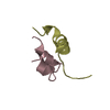
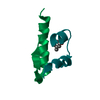
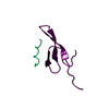
| Title | Crystal structure of HERC2 ZZ domain in complex with SUMO1 tail | ||||||
 Components Components | SUMO1 linked HERC2 ZZ domain (Small ubiquitin-related modifier 1,E3 ubiquitin-protein ligase HERC2) | ||||||
 Keywords Keywords |  GENE REGULATION / Zn finger protein / ZZ domain / HERC2 GENE REGULATION / Zn finger protein / ZZ domain / HERC2 | ||||||
| Function / homology |  Function and homology information Function and homology informationnegative regulation of transcription by transcription factor localization / SUMOylation of nuclear envelope proteins / Negative regulation of activity of TFAP2 (AP-2) family transcription factors / SUMO is proteolytically processed / negative regulation of delayed rectifier potassium channel activity / SUMO is conjugated to E1 (UBA2:SAE1) / nuclear stress granule / SUMO is transferred from E1 to E2 (UBE2I, UBC9) / negative regulation of action potential / SUMO binding ...negative regulation of transcription by transcription factor localization / SUMOylation of nuclear envelope proteins / Negative regulation of activity of TFAP2 (AP-2) family transcription factors / SUMO is proteolytically processed / negative regulation of delayed rectifier potassium channel activity / SUMO is conjugated to E1 (UBA2:SAE1) / nuclear stress granule / SUMO is transferred from E1 to E2 (UBE2I, UBC9) / negative regulation of action potential / SUMO binding / small protein activating enzyme binding / SUMOylation of DNA methylation proteins / HECT-type E3 ubiquitin transferase / SUMOylation of immune response proteins / SUMOylation of SUMOylation proteins / Maturation of nucleoprotein / SUMOylation of RNA binding proteins / Postmitotic nuclear pore complex (NPC) reformation / Maturation of nucleoprotein / negative regulation of protein import into nucleus / ubiquitin-specific protease binding / roof of mouth development / SUMOylation of ubiquitinylation proteins / negative regulation of DNA binding / ubiquitin-like protein ligase binding / SUMOylation of DNA replication proteins / protein sumoylation / transcription factor binding / SUMOylation of transcription factors / potassium channel regulator activity / SUMOylation of DNA damage response and repair proteins / Regulation of IFNG signaling / nuclear pore / cellular response to cadmium ion / centriole / SUMOylation of chromatin organization proteins / guanyl-nucleotide exchange factor activity / SUMOylation of transcription cofactors / positive regulation of protein-containing complex assembly / Nonhomologous End-Joining (NHEJ) / intracellular protein transport / SUMOylation of intracellular receptors / PKR-mediated signaling / G2/M DNA damage checkpoint / negative regulation of DNA-binding transcription factor activity / PML body / protein tag activity / Formation of Incision Complex in GG-NER / regulation of protein localization / ubiquitin protein ligase activity / positive regulation of proteasomal ubiquitin-dependent protein catabolic process / Antigen processing: Ubiquitination & Proteasome degradation / Recruitment and ATM-mediated phosphorylation of repair and signaling proteins at DNA double strand breaks / cellular response to heat / Processing of DNA double-strand break ends / spermatogenesis / proteasome-mediated ubiquitin-dependent protein catabolic process / nuclear membrane / protein stabilization / protein ubiquitination / nuclear body / nuclear speck / DNA repair / negative regulation of DNA-templated transcription / DNA damage response / ubiquitin protein ligase binding / nucleolus / enzyme binding / RNA binding / zinc ion binding / nucleoplasm / membrane / nucleus / plasma membrane / cytosol / cytoplasmSimilarity search - Function | ||||||
| Biological species |  Homo sapiens (human) Homo sapiens (human) | ||||||
| Method | X-RAY DIFFRACTION / MOLECULAR REPLACEMENT / Resolution: 2.096 Å | ||||||
 Authors Authors | Liu, J. / Vann, K.R. / Kutateladze, T.G. | ||||||
 Citation Citation | Journal: Structure / Year: 2020 Title: Structural Insight into Binding of the ZZ Domain of HERC2 to Histone H3 and SUMO1. Authors: Liu, J. / Xue, Z. / Zhang, Y. / Vann, K.R. / Shi, X. / Kutateladze, T.G. | ||||||
| History |
|
- Structure visualization
| Structure viewer | Molecule: MolmilJmol/JSmol |
|---|
- Downloads & links
-Download
| PDBx/mmCIF format | 6ww3.cif.gz | 42.6 KB | Display | PDBx/mmCIF format |
|---|---|---|---|---|
| PDB format | pdb6ww3.ent.gz | 27.3 KB | Display | PDB format |
| PDBx/mmJSON format | 6ww3.json.gz | Tree view | PDBx/mmJSON format | |
| Others |  Other downloads Other downloads |
-Validation report
| Arichive directory | https://data.pdbj.org/pub/pdb/validation_reports/ww/6ww3ftp://data.pdbj.org/pub/pdb/validation_reports/ww/6ww3 | HTTPS FTP |
|---|
-Related structure data
| Related structure data |  6ww4C  6ds6S S: Starting model for refinement C: citing same article ( |
|---|---|
| Similar structure data |


-Links
 PDBj
PDBj


- Assembly
| Deposited unit | 
| ||||||||
|---|---|---|---|---|---|---|---|---|---|
| 1 | 
| ||||||||
| 2 | 
| ||||||||
| Unit cell |
|
-Components
| #1: Protein | Mass: 6909.763 Da / Num. of mol.: 2 / Fragment: fusion protein Source method: isolated from a genetically manipulated source Source: (gene. exp.) Homo sapiens (human) / Gene: SUMO1, SMT3C, SMT3H3, UBL1, OK/SW-cl.43, HERC2 / Production host:  Escherichia coli (E. coli) Escherichia coli (E. coli)References: UniProt: P63165, UniProt: O95714, HECT-type E3 ubiquitin transferase #2: Chemical | ChemComp-ZN /   Mass: 65.409 Da / Num. of mol.: 4 / Source method: obtained synthetically / Formula: Zn Mass: 65.409 Da / Num. of mol.: 4 / Source method: obtained synthetically / Formula: Zn#3: Chemical | ChemComp-PEG / | Diethylene glycol  Mass: 106.120 Da / Num. of mol.: 1 / Source method: obtained synthetically / Formula: C4H10O3 Mass: 106.120 Da / Num. of mol.: 1 / Source method: obtained synthetically / Formula: C4H10O3#4: Chemical | ChemComp-GOL / | Glycerol  Mass: 92.094 Da / Num. of mol.: 1 / Source method: obtained synthetically / Formula: C3H8O3 Mass: 92.094 Da / Num. of mol.: 1 / Source method: obtained synthetically / Formula: C3H8O3#5: Water | ChemComp-HOH / | Water Mass: 18.015 Da / Num. of mol.: 129 / Source method: isolated from a natural source / Formula: H2O Mass: 18.015 Da / Num. of mol.: 129 / Source method: isolated from a natural source / Formula: H2OHas ligand of interest | N | Sequence details | Residues 2-7 of SUMO1 fused to the ZZ domain of HERC2 | |
|---|
-Experimental details
-Experiment
| Experiment | Method: X-RAY DIFFRACTION / Number of used crystals: 1 |
|---|
- Sample preparation
| Crystal | Density Matthews: 1.99 Å3/Da / Density % sol: 38.08 % |
|---|---|
| Crystal grow | Temperature: 298 K / Method: vapor diffusion, sitting drop Details: 0.2 M ammonium chloride, 0.1 M Tris, pH 8.0 and 20% PEG 6000 |
-Data collection
| Diffraction | Mean temperature: 100 K / Serial crystal experiment: N | |||||||||||||||||||||||||||||||||||||||||||||||||||||||||||||||||||||||||||||||||||||||||||||||||||
|---|---|---|---|---|---|---|---|---|---|---|---|---|---|---|---|---|---|---|---|---|---|---|---|---|---|---|---|---|---|---|---|---|---|---|---|---|---|---|---|---|---|---|---|---|---|---|---|---|---|---|---|---|---|---|---|---|---|---|---|---|---|---|---|---|---|---|---|---|---|---|---|---|---|---|---|---|---|---|---|---|---|---|---|---|---|---|---|---|---|---|---|---|---|---|---|---|---|---|---|---|
| Diffraction source | Source: ROTATING ANODE / Type: RIGAKU MICROMAX-007 HF / Wavelength: 1.54 Å | |||||||||||||||||||||||||||||||||||||||||||||||||||||||||||||||||||||||||||||||||||||||||||||||||||
| Detector | Type: DECTRIS PILATUS 200K / Detector: PIXEL / Date: Oct 22, 2018 | |||||||||||||||||||||||||||||||||||||||||||||||||||||||||||||||||||||||||||||||||||||||||||||||||||
| Radiation | Protocol: SINGLE WAVELENGTH / Monochromatic (M) / Laue (L): M / Scattering type: x-ray | |||||||||||||||||||||||||||||||||||||||||||||||||||||||||||||||||||||||||||||||||||||||||||||||||||
| Radiation wavelength | Wavelength: 1.54 Å / Relative weight: 1 | |||||||||||||||||||||||||||||||||||||||||||||||||||||||||||||||||||||||||||||||||||||||||||||||||||
| Reflection | Resolution: 1.95→50 Å / Num. obs: 7588 / % possible obs: 89.2 % / Redundancy: 3 % / Biso Wilson estimate: 13.18 Å2 / Rmerge(I) obs: 0.181 / Rpim(I) all: 0.113 / Rrim(I) all: 0.215 / Χ2: 1.062 / Net I/σ(I): 3.5 / Num. measured all: 22967 | |||||||||||||||||||||||||||||||||||||||||||||||||||||||||||||||||||||||||||||||||||||||||||||||||||
| Reflection shell | Diffraction-ID: 1
|
- Processing
| Software |
| ||||||||||||||||||||||||
|---|---|---|---|---|---|---|---|---|---|---|---|---|---|---|---|---|---|---|---|---|---|---|---|---|---|
| Refinement | Method to determine structure: MOLECULAR REPLACEMENT Starting model: 6DS6 Resolution: 2.096→33.426 Å / SU ML: 0.19 / Cross valid method: FREE R-VALUE / σ(F): 1.34 / Phase error: 21.17 / Stereochemistry target values: ML
| ||||||||||||||||||||||||
| Solvent computation | Shrinkage radii: 0.9 Å / VDW probe radii: 1.11 Å / Solvent model: FLAT BULK SOLVENT MODEL | ||||||||||||||||||||||||
| Displacement parameters | Biso max: 42.5 Å2 / Biso mean: 14.5705 Å2 / Biso min: 4.74 Å2 | ||||||||||||||||||||||||
| Refinement step | Cycle: final / Resolution: 2.096→33.426 Å
| ||||||||||||||||||||||||
| Refine LS restraints |
| ||||||||||||||||||||||||
| LS refinement shell | Refine-ID: X-RAY DIFFRACTION / Rfactor Rfree error: 0
|
