 Movie
Movie Controller


+ Open data

- Basic information
| Entry | Database: PDB / ID: 2lvn | ||||||
|---|---|---|---|---|---|---|---|
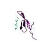
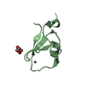
| Title | Structure of the gp78 CUE domain | ||||||
 Components Components | E3 ubiquitin-protein ligase AMFR | ||||||
 Keywords Keywords |  LIGASE / CUE domain LIGASE / CUE domain | ||||||
| Function / homology |  Function and homology information Function and homology informationregulation of SREBP signaling pathway / positive regulation of plant-type hypersensitive response / RING-type E3 ubiquitin transferase (cysteine targeting) / protein K27-linked ubiquitination / Derlin-1 retrotranslocation complex / BAT3 complex binding / ubiquitin-ubiquitin ligase activity / ubiquitin-specific protease binding / ERAD pathway / non-canonical NF-kappaB signal transduction ...regulation of SREBP signaling pathway / positive regulation of plant-type hypersensitive response / RING-type E3 ubiquitin transferase (cysteine targeting) / protein K27-linked ubiquitination / Derlin-1 retrotranslocation complex / BAT3 complex binding / ubiquitin-ubiquitin ligase activity / ubiquitin-specific protease binding / ERAD pathway / non-canonical NF-kappaB signal transduction / protein autoubiquitination / protein K48-linked ubiquitination / ubiquitin ligase complex / endoplasmic reticulum unfolded protein response / ER Quality Control Compartment (ERQC) / N-glycan trimming in the ER and Calnexin/Calreticulin cycle / ubiquitin binding / negative regulation of canonical Wnt signaling pathway / Wnt signaling pathway / protein polyubiquitination / ubiquitin-protein transferase activity / ubiquitin protein ligase activity / protein-macromolecule adaptor activity / signaling receptor activity / protein-folding chaperone binding / growth cone / ubiquitin-dependent protein catabolic process / learning or memory / neuronal cell body / dendrite / endoplasmic reticulum membrane / perinuclear region of cytoplasm / Golgi apparatus / endoplasmic reticulum / signal transduction / protein-containing complex / membrane / identical protein binding / metal ion binding / plasma membrane / cytosolSimilarity search - Function | ||||||
| Biological species |  Homo sapiens (human) Homo sapiens (human) | ||||||
| Method | SOLUTION NMR / simulated annealing | ||||||
| Model details | lowest energy, model 1 | ||||||
 Authors Authors | Liu, S. / Chen, Y. / Huang, T. / Tarasov, S.G. / King, A. / Li, J. / Weissman, A.M. / Byrd, R.A. / Das, R. | ||||||
 Citation Citation | Journal: Structure / Year: 2012 Title: Promiscuous Interactions of gp78 E3 Ligase CUE Domain with Polyubiquitin Chains. Authors: Liu, S. / Chen, Y. / Li, J. / Huang, T. / Tarasov, S. / King, A. / Weissman, A.M. / Byrd, R.A. / Das, R. | ||||||
| History |
|
- Structure visualization
| Structure viewer | Molecule: MolmilJmol/JSmol |
|---|
- Downloads & links
-Download
| PDBx/mmCIF format | 2lvn.cif.gz | 329.6 KB | Display | PDBx/mmCIF format |
|---|---|---|---|---|
| PDB format | pdb2lvn.ent.gz | 275.6 KB | Display | PDB format |
| PDBx/mmJSON format | 2lvn.json.gz | Tree view | PDBx/mmJSON format | |
| Others |  Other downloads Other downloads |
-Validation report
| Arichive directory | https://data.pdbj.org/pub/pdb/validation_reports/lv/2lvnftp://data.pdbj.org/pub/pdb/validation_reports/lv/2lvn | HTTPS FTP |
|---|
-Related structure data
| Related structure data |  2lvoC  2lvpC  2lvqC C: citing same article ( |
|---|---|
| Similar structure data | |
| Other databases |


-Links
 PDBj
PDBj


- Assembly
| Deposited unit | 
| |||||||||
|---|---|---|---|---|---|---|---|---|---|---|
| 1 |
| |||||||||
| NMR ensembles |
|
-Components
| #1: Protein | Mass: 5966.752 Da / Num. of mol.: 1 / Fragment: CUE domain residues 453-504 Source method: isolated from a genetically manipulated source Source: (gene. exp.) Homo sapiens (human) / Gene: AMFR, RNF45 / Production host:  Escherichia coli (E. coli) Escherichia coli (E. coli)References: UniProt: Q9UKV5, Ligases; Forming carbon-nitrogen bonds; Acid-amino-acid ligases (peptide synthases) |
|---|
-Experimental details
-Experiment
| Experiment | Method: SOLUTION NMR / Details: 1H, 13C, 15N assignments of gp78 CUE domain | ||||||||||||||||||||||||||||||||||||||||
|---|---|---|---|---|---|---|---|---|---|---|---|---|---|---|---|---|---|---|---|---|---|---|---|---|---|---|---|---|---|---|---|---|---|---|---|---|---|---|---|---|---|
| NMR experiment |
|
- Sample preparation
| Details | Contents: 50 mM TRIS, 50 mM sodium chloride, 90% H2O/10% D2O Solvent system: 90% H2O/10% D2O | |||||||||
|---|---|---|---|---|---|---|---|---|---|---|
| Sample |
| |||||||||
| Sample conditions | pH: 7.2 / Pressure: ambient / Temperature: 298 K |
-NMR measurement
| NMR spectrometer |
|
|---|
- Processing
| NMR software |
| ||||||||||||||||||
|---|---|---|---|---|---|---|---|---|---|---|---|---|---|---|---|---|---|---|---|
| Refinement | Method: simulated annealing / Software ordinal: 1 / Details: FINAL STEP IN WATER | ||||||||||||||||||
| NMR constraints | NOE constraints total: 1189 / NOE intraresidue total count: 282 / NOE long range total count: 102 / NOE medium range total count: 634 / NOE sequential total count: 171 / Protein phi angle constraints total count: 48 / Protein psi angle constraints total count: 49 | ||||||||||||||||||
| NMR representative | Selection criteria: lowest energy | ||||||||||||||||||
| NMR ensemble | Conformer selection criteria: structures with the lowest energy Conformers calculated total number: 100 / Conformers submitted total number: 20 / Maximum lower distance constraint violation: 1.8 Å / Maximum upper distance constraint violation: 6 Å | ||||||||||||||||||
| NMR ensemble rms | Distance rms dev: 0 Å |
