| Entry | Database: PDB / ID: 6wte
|
|---|
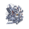
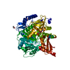
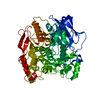
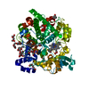
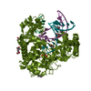
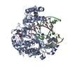
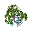
| Title | Structure of radical S-adenosylmethionine methyltransferase, TsrM, from Kitasatospora setae with cobalamin and [4Fe-4S] cluster bound |
|---|
 Components Components | B12-binding domain-containing protein |
|---|
 Keywords Keywords |  TRANSFERASE / Radical SAM / Cobalamin / FeS cluster / methyltransferase TRANSFERASE / Radical SAM / Cobalamin / FeS cluster / methyltransferase |
|---|
| Function / homology |  Function and homology information Function and homology information |
|---|
| Biological species |  Kitasatospora setae (bacteria) Kitasatospora setae (bacteria) |
|---|
| Method | X-RAY DIFFRACTION / SYNCHROTRON / SAD / Resolution: 1.67 Å |
|---|
 Authors Authors | Knox, H.L. / Chen, P.Y.-T. / Drennan, C.L. / Booker, S.J. |
|---|
| Funding support |  United States, 2items United States, 2items | Organization | Grant number | Country |
|---|
| National Institutes of Health/National Institute of General Medical Sciences (NIH/NIGMS) | GM-12259 | United States | | National Institutes of Health/National Institute of General Medical Sciences (NIH/NIGMS) | GM-126982 | United States |
|
|---|
 Citation Citation | Journal: Nat.Chem.Biol. / Year: 2021
Title: Structural basis for non-radical catalysis by TsrM, a radical SAM methylase.
Authors: Knox, H.L. / Chen, P.Y. / Blaszczyk, A.J. / Mukherjee, A. / Grove, T.L. / Schwalm, E.L. / Wang, B. / Drennan, C.L. / Booker, S.J. |
|---|
| History | | Deposition | May 2, 2020 | Deposition site: RCSB / Processing site: RCSB |
|---|
| Revision 1.0 | Dec 23, 2020 | Provider: repository / Type: Initial release |
|---|
| Revision 1.1 | Feb 3, 2021 | Group: Database references / Category: citation / citation_author
Item: _citation.pdbx_database_id_PubMed / _citation.title ..._citation.pdbx_database_id_PubMed / _citation.title / _citation.year / _citation_author.name |
|---|
| Revision 1.2 | Apr 7, 2021 | Group: Database references / Category: citation
Item: _citation.journal_volume / _citation.page_first / _citation.page_last |
|---|
| Revision 1.3 | Mar 6, 2024 | Group: Data collection / Database references / Category: chem_comp_atom / chem_comp_bond / database_2
Item: _database_2.pdbx_DOI / _database_2.pdbx_database_accession |
|---|
|
|---|
 Movie
Movie Controller

 Yorodumi
 Open data
 Basic information
 Structure visualization
 Downloads & links
 Other downloads
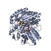


 PDBj
PDBj Assembly