 Movie
Movie Controller

[English] 日本語
 Yorodumi
- PDB-6wt9: Structure of STING-associated CdnE c-di-GMP synthase from Capnocy... -
+ Open data

- Basic information
| Entry | Database: PDB / ID: 6wt9 | ||||||
|---|---|---|---|---|---|---|---|
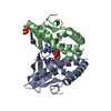
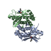
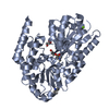
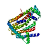
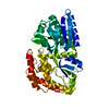
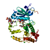
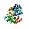
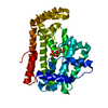
| Title | Structure of STING-associated CdnE c-di-GMP synthase from Capnocytophaga granulosa | ||||||
 Components Components | NTP_transf_2 domain-containing protein | ||||||
 Keywords Keywords | TRANSFERASE / CBASS / CD-NTase / c-di-GMP / STING | ||||||
| Function / homology | diguanylate cyclase / diguanylate cyclase activity / Polymerase, nucleotidyl transferase domain / Nucleotidyltransferase domain / Nucleotidyltransferase superfamily / defense response to virus / GTP binding / c-di-GMP synthase Function and homology information Function and homology information | ||||||
| Biological species |  Capnocytophaga granulosa (bacteria) Capnocytophaga granulosa (bacteria) | ||||||
| Method |  X-RAY DIFFRACTION / SYNCHROTRON / SAD / Resolution: 2.3 Å X-RAY DIFFRACTION / SYNCHROTRON / SAD / Resolution: 2.3 Å | ||||||
 Authors Authors | Morehouse, B.R. / Govande, A.A. / Millman, A. / Keszei, A.F.A. / Lowey, B. / Ofir, G. / Shao, S. / Sorek, R. / Kranzusch, P.J. | ||||||
 Citation Citation | Journal: Nature / Year: 2020 Title: STING cyclic dinucleotide sensing originated in bacteria. Authors: Morehouse, B.R. / Govande, A.A. / Millman, A. / Keszei, A.F.A. / Lowey, B. / Ofir, G. / Shao, S. / Sorek, R. / Kranzusch, P.J. | ||||||
| History |
|
- Structure visualization
| Structure viewer | Molecule: MolmilJmol/JSmol |
|---|
- Downloads & links
-Download
| PDBx/mmCIF format | 6wt9.cif.gz | 161 KB | Display | PDBx/mmCIF format |
|---|---|---|---|---|
| PDB format | pdb6wt9.ent.gz | 110.7 KB | Display | PDB format |
| PDBx/mmJSON format | 6wt9.json.gz | Tree view | PDBx/mmJSON format | |
| Others |  Other downloads Other downloads |
-Validation report
| Summary document | 6wt9_validation.pdf.gz | 421.5 KB | Display | wwPDB validaton report |
|---|---|---|---|---|
| Full document | 6wt9_full_validation.pdf.gz | 423.8 KB | Display | |
| Data in XML | 6wt9_validation.xml.gz | 14 KB | Display | |
| Data in CIF | 6wt9_validation.cif.gz | 19.7 KB | Display | |
| Arichive directory | https://data.pdbj.org/pub/pdb/validation_reports/wt/6wt9ftp://data.pdbj.org/pub/pdb/validation_reports/wt/6wt9 | HTTPS FTP |
-Related structure data


-Links
 PDBj
PDBj
- Assembly
| Deposited unit | 
| ||||||||||||
|---|---|---|---|---|---|---|---|---|---|---|---|---|---|
| 1 |
| ||||||||||||
| Unit cell |
|
-Components
| #1: Protein | Mass: 41378.113 Da / Num. of mol.: 1 Source method: isolated from a genetically manipulated source Source: (gene. exp.) Capnocytophaga granulosa (bacteria) / Gene: NCTC12948_02564 / Production host: |
|---|---|
| #2: Water | ChemComp-HOH /  Mass: 18.015 Da / Num. of mol.: 169 / Source method: isolated from a natural source / Formula: H2O Mass: 18.015 Da / Num. of mol.: 169 / Source method: isolated from a natural source / Formula: H2O |
| Has ligand of interest | N |
-Experimental details
-Experiment
| Experiment | Method: X-RAY DIFFRACTION / Number of used crystals: 1 |
|---|
- Sample preparation
| Crystal | Density Matthews: 1.99 Å3/Da / Density % sol: 38.2 % |
|---|---|
| Crystal grow | Temperature: 291 K / Method: vapor diffusion, hanging drop / Details: 0.2 M sodium thiocyanate, 20% PEG-3350 |
-Data collection
| Diffraction | Mean temperature: 80 K / Serial crystal experiment: N |
|---|---|
| Diffraction source | Source: SYNCHROTRON / Site: APS  / Beamline: 24-ID-E / Wavelength: 0.97918 Å / Beamline: 24-ID-E / Wavelength: 0.97918 Å |
| Detector | Type: DECTRIS EIGER X 16M / Detector: PIXEL / Date: Dec 12, 2019 |
| Radiation | Protocol: SINGLE WAVELENGTH / Monochromatic (M) / Laue (L): M / Scattering type: x-ray |
| Radiation wavelength | Wavelength: 0.97918 Å / Relative weight: 1 |
| Reflection | Resolution: 2.3→44.85 Å / Num. obs: 14608 / % possible obs: 99.6 % / Redundancy: 10.4 % / Biso Wilson estimate: 18.54 Å2 / CC1/2: 0.961 / Rpim(I) all: 0.096 / Net I/σ(I): 17 |
| Reflection shell | Resolution: 2.3→2.38 Å / Num. unique obs: 1408 / CC1/2: 0.769 / Rpim(I) all: 0.457 |
- Processing
| Software |
| ||||||||||||||||||||||||||||||||||||||||||||||||||||||||||||||||||||||||||||||||||||||||||||||||||||||||||||||||||||||||||||||||||||||||||||||||||||||
|---|---|---|---|---|---|---|---|---|---|---|---|---|---|---|---|---|---|---|---|---|---|---|---|---|---|---|---|---|---|---|---|---|---|---|---|---|---|---|---|---|---|---|---|---|---|---|---|---|---|---|---|---|---|---|---|---|---|---|---|---|---|---|---|---|---|---|---|---|---|---|---|---|---|---|---|---|---|---|---|---|---|---|---|---|---|---|---|---|---|---|---|---|---|---|---|---|---|---|---|---|---|---|---|---|---|---|---|---|---|---|---|---|---|---|---|---|---|---|---|---|---|---|---|---|---|---|---|---|---|---|---|---|---|---|---|---|---|---|---|---|---|---|---|---|---|---|---|---|---|---|---|
| Refinement | Method to determine structure: SAD / Resolution: 2.3→44.85 Å / SU ML: 0.2445 / Cross valid method: FREE R-VALUE / σ(F): 1.34 / Phase error: 24.6953
| ||||||||||||||||||||||||||||||||||||||||||||||||||||||||||||||||||||||||||||||||||||||||||||||||||||||||||||||||||||||||||||||||||||||||||||||||||||||
| Solvent computation | Shrinkage radii: 0.9 Å / VDW probe radii: 1.11 Å | ||||||||||||||||||||||||||||||||||||||||||||||||||||||||||||||||||||||||||||||||||||||||||||||||||||||||||||||||||||||||||||||||||||||||||||||||||||||
| Displacement parameters | Biso mean: 28.12 Å2 | ||||||||||||||||||||||||||||||||||||||||||||||||||||||||||||||||||||||||||||||||||||||||||||||||||||||||||||||||||||||||||||||||||||||||||||||||||||||
| Refinement step | Cycle: LAST / Resolution: 2.3→44.85 Å
| ||||||||||||||||||||||||||||||||||||||||||||||||||||||||||||||||||||||||||||||||||||||||||||||||||||||||||||||||||||||||||||||||||||||||||||||||||||||
| Refine LS restraints |
| ||||||||||||||||||||||||||||||||||||||||||||||||||||||||||||||||||||||||||||||||||||||||||||||||||||||||||||||||||||||||||||||||||||||||||||||||||||||
| LS refinement shell |
| ||||||||||||||||||||||||||||||||||||||||||||||||||||||||||||||||||||||||||||||||||||||||||||||||||||||||||||||||||||||||||||||||||||||||||||||||||||||
| Refinement TLS params. | Method: refined / Refine-ID: X-RAY DIFFRACTION
| ||||||||||||||||||||||||||||||||||||||||||||||||||||||||||||||||||||||||||||||||||||||||||||||||||||||||||||||||||||||||||||||||||||||||||||||||||||||
| Refinement TLS group |
|
