[English] 日本語
Yorodumi
- PDB-6wle: Crystal structure of the Zeitlupe light-state mimic G46A -

+
Open data


ID or keywords:

Loading...

-
Basic information

Entry
Database: PDB / ID: 6wle
TitleCrystal structure of the Zeitlupe light-state mimic G46A
ComponentsAdagio protein 1
KeywordsPLANT PROTEIN / Circadian clock control / dimer
Function / homology
Function and homology information


response to blue light / flower development / SCF ubiquitin ligase complex / photoreceptor activity / circadian rhythm / protein ubiquitination / nucleus / cytosol
Similarity search - Function
Kelch repeat type 2 / Kelch motif / Galactose oxidase, central domain / Galactose oxidase, central domain / A Receptor for Ubiquitination Targets / F-box domain profile. / Galactose oxidase/kelch, beta-propeller / F-box-like domain superfamily / F-box-like / F-box domain ...Kelch repeat type 2 / Kelch motif / Galactose oxidase, central domain / Galactose oxidase, central domain / A Receptor for Ubiquitination Targets / F-box domain profile. / Galactose oxidase/kelch, beta-propeller / F-box-like domain superfamily / F-box-like / F-box domain / PAS domain / Kelch-type beta propeller / PAS repeat profile. / PAS domain / PAS domain superfamily
Similarity search - Domain/homology
FLAVIN MONONUCLEOTIDE / Adagio protein 1
Similarity search - Component
Biological speciesArabidopsis thaliana (thale cress)
MethodX-RAY DIFFRACTION / SYNCHROTRON / MOLECULAR REPLACEMENT / Resolution: 3 Å
AuthorsZoltowski, B. / Green, R.
Funding support United States, 1items
OrganizationGrant numberCountry
National Institutes of Health/National Institute of General Medical Sciences (NIH/NIGMS)2R15GM109282 United States
CitationJournal: Biochemistry / Year: 2021
Title: Steric and Electronic Interactions at Gln154 in ZEITLUPE Induce Reorganization of the LOV Domain Dimer Interface.
Authors: Pudasaini, A. / Green, R. / Song, Y.H. / Blumenfeld, A. / Karki, N. / Imaizumi, T. / Zoltowski, B.D.
History
DepositionApr 20, 2020Deposition site: RCSB / Processing site: RCSB
Revision 1.0Mar 3, 2021Provider: repository / Type: Initial release
Revision 1.1Oct 18, 2023Group: Data collection / Database references / Refinement description
Category: chem_comp_atom / chem_comp_bond ...chem_comp_atom / chem_comp_bond / database_2 / pdbx_initial_refinement_model
Item: _database_2.pdbx_DOI / _database_2.pdbx_database_accession

-
Structure visualization

Structure viewerMolecule:
MolmilJmol/JSmol

Downloads & links

-
Assembly

Deposited unit
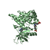
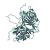
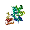
B: Adagio protein 1
A: Adagio protein 1
C: Adagio protein 1
D: Adagio protein 1
E: Adagio protein 1
F: Adagio protein 1
G: Adagio protein 1
hetero molecules


Theoretical massNumber of molelcules
Total (without water)149,08817
Polymers145,7087
Non-polymers3,38110
Water1,838102
1
B: Adagio protein 1
D: Adagio protein 1
hetero molecules


Theoretical massNumber of molelcules
Total (without water)42,6065
Polymers41,6312
Non-polymers9753
Water362
TypeNameSymmetry operationNumber
identity operation1_555x,y,z1
Buried area3820 Å2
ΔGint-23 kcal/mol
Surface area14260 Å2
MethodPISA
2
A: Adagio protein 1
E: Adagio protein 1
hetero molecules


Theoretical massNumber of molelcules
Total (without water)42,6065
Polymers41,6312
Non-polymers9753
Water362
TypeNameSymmetry operationNumber
identity operation1_555x,y,z1
Buried area3820 Å2
ΔGint-23 kcal/mol
Surface area14290 Å2
MethodPISA
3
C: Adagio protein 1
G: Adagio protein 1
hetero molecules


Theoretical massNumber of molelcules
Total (without water)42,6065
Polymers41,6312
Non-polymers9753
Water362
TypeNameSymmetry operationNumber
identity operation1_555x,y,z1
Buried area3960 Å2
ΔGint-21 kcal/mol
Surface area14190 Å2
MethodPISA
4
F: Adagio protein 1
hetero molecules

F: Adagio protein 1
hetero molecules


Theoretical massNumber of molelcules
Total (without water)42,5434
Polymers41,6312
Non-polymers9132
Water362
TypeNameSymmetry operationNumber
identity operation1_555x,y,z1
crystal symmetry operation15_455-x-1/2,y,-z1
Buried area3740 Å2
ΔGint-26 kcal/mol
Surface area14500 Å2
MethodPISA
Unit cell
Length a, b, c (Å)265.822, 265.822, 265.822
Angle α, β, γ (deg.)90.000, 90.000, 90.000
Int Tables number199
Space group name H-MI213
Space group name HallI2b2c3
Symmetry operation#1: x,y,z
#2: z,x,y
#3: y,z,x
#4: -y,-z+1/2,x
#5: z,-x,-y+1/2
#6: -y+1/2,z,-x
#7: -z,-x+1/2,y
#8: -z+1/2,x,-y
#9: y,-z,-x+1/2
#10: x,-y,-z+1/2
#11: -x+1/2,y,-z
#12: -x,-y+1/2,z
#13: x+1/2,y+1/2,z+1/2
#14: z+1/2,x+1/2,y+1/2
#15: y+1/2,z+1/2,x+1/2
#16: -y+1/2,-z+1,x+1/2
#17: z+1/2,-x+1/2,-y+1
#18: -y+1,z+1/2,-x+1/2
#19: -z+1/2,-x+1,y+1/2
#20: -z+1,x+1/2,-y+1/2
#21: y+1/2,-z+1/2,-x+1
#22: x+1/2,-y+1/2,-z+1
#23: -x+1,y+1/2,-z+1/2
#24: -x+1/2,-y+1,z+1/2
Components on special symmetry positions
IDModelComponents
11F-17-

CYS

-
Components

#1: Protein
Adagio protein 1 / Clock-associated PAS protein ZTL / F-box only protein 2b / FBX2b / Flavin-binding kelch repeat F- ...Clock-associated PAS protein ZTL / F-box only protein 2b / FBX2b / Flavin-binding kelch repeat F-box protein 1-like protein 2 / FKF1-like protein 2 / LOV kelch protein 1 / Protein ZEITLUPE


Mass: 20815.404 Da / Num. of mol.: 7 / Mutation: G46A
Source method: isolated from a genetically manipulated source
Source: (gene. exp.) Arabidopsis thaliana (thale cress) / Gene: ADO1, FKL2, LKP1, ZTL, At5g57360, MSF19.2 / Production host: Escherichia coli (E. coli) / References: UniProt: Q94BT6
#2: Chemical
ChemComp-FMN / FLAVIN MONONUCLEOTIDE / RIBOFLAVIN MONOPHOSPHATE / Flavin mononucleotide


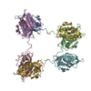
Mass: 456.344 Da / Num. of mol.: 7 / Source method: obtained synthetically / Formula: C17H21N4O9P
#3: Chemical ChemComp-EDO / 1,2-ETHANEDIOL / ETHYLENE GLYCOL / Ethylene glycol


Mass: 62.068 Da / Num. of mol.: 3 / Source method: obtained synthetically / Formula: C2H6O2
#4: Water ChemComp-HOH / water / Water


Mass: 18.015 Da / Num. of mol.: 102 / Source method: isolated from a natural source / Formula: H2O
Has ligand of interestN

-
Experimental details

-
Experiment

ExperimentMethod: X-RAY DIFFRACTION / Number of used crystals: 1

-
Sample preparation

CrystalDensity Matthews: 5.37 Å3/Da / Density % sol: 77.1 %
Crystal growTemperature: 293 K / Method: vapor diffusion, hanging drop / pH: 6
Details: 0.1 M MES pH 6.0, 1.6 M Ammonium Sulfate, 0.01 M Cobalt Chloride Hexahydrate

-
Data collection

DiffractionMean temperature: 80 K / Serial crystal experiment: N
Diffraction sourceSource: SYNCHROTRON / Site: CHESS / Beamline: F1 / Wavelength: 0.9773 Å
DetectorType: DECTRIS PILATUS3 6M / Detector: PIXEL / Date: Nov 17, 2016
RadiationProtocol: SINGLE WAVELENGTH / Monochromatic (M) / Laue (L): M / Scattering type: x-ray
Radiation wavelengthWavelength: 0.9773 Å / Relative weight: 1
ReflectionResolution: 3→45.59 Å / Num. obs: 62237 / % possible obs: 99.7 % / Redundancy: 6.4 % / Biso Wilson estimate: 53.42 Å2 / Rmerge(I) obs: 0.172 / Net I/σ(I): 17.5
Reflection shellResolution: 3→3.05 Å / Num. unique obs: 3079 / CC1/2: 0.642

-
Processing

Software
NameVersionClassification
PHENIX1.17.1_3660refinement
HKL-2000data reduction
HKL-2000data scaling
PHENIXphasing
RefinementMethod to determine structure: MOLECULAR REPLACEMENT
Starting model: 5SVU
Resolution: 3→45.59 Å / SU ML: 0.396 / Cross valid method: FREE R-VALUE / σ(F): 1.34 / Phase error: 25.1092
RfactorNum. reflection% reflection
Rfree0.2425 1997 3.21 %
Rwork0.2029 --
obs0.2041 62142 99.79 %
Solvent computationShrinkage radii: 0.9 Å / VDW probe radii: 1.11 Å
Displacement parametersBiso mean: 48.57 Å2
Refinement stepCycle: LAST / Resolution: 3→45.59 Å
ProteinNucleic acidLigandSolventTotal
Num. atoms7243 0 229 102 7574
Refine LS restraints
Refine-IDTypeDev idealNumber
X-RAY DIFFRACTIONf_bond_d0.01737631
X-RAY DIFFRACTIONf_angle_d1.558710375
X-RAY DIFFRACTIONf_chiral_restr0.07011157
X-RAY DIFFRACTIONf_plane_restr0.00891328
X-RAY DIFFRACTIONf_dihedral_angle_d18.42171049
LS refinement shell
Resolution (Å)Rfactor RfreeNum. reflection RfreeRfactor RworkNum. reflection RworkRefine-ID% reflection obs (%)
3-3.080.32341400.29694283X-RAY DIFFRACTION99.93
3.08-3.160.34581440.28514275X-RAY DIFFRACTION99.86
3.16-3.250.32311440.27064277X-RAY DIFFRACTION99.77
3.25-3.360.32991450.25874264X-RAY DIFFRACTION99.95
3.36-3.480.2841380.26964268X-RAY DIFFRACTION99.86
3.48-3.610.27671450.21864267X-RAY DIFFRACTION99.91
3.62-3.780.27371460.25724237X-RAY DIFFRACTION99.3
3.78-3.980.31021390.25434274X-RAY DIFFRACTION99.59
3.98-4.230.21031440.17244307X-RAY DIFFRACTION99.98
4.23-4.550.20071400.13824282X-RAY DIFFRACTION100
4.55-5.010.14551420.13064340X-RAY DIFFRACTION100
5.01-5.740.16841450.14894313X-RAY DIFFRACTION100
5.74-7.220.20541400.17874348X-RAY DIFFRACTION100
7.22-45.590.24221450.19384410X-RAY DIFFRACTION98.89

+
About Yorodumi

-
News

-
Feb 9, 2022. New format data for meta-information of EMDB entries

New format data for meta-information of EMDB entries

  • Version 3 of the EMDB header file is now the official format.
  • The previous official version 1.9 will be removed from the archive.

Related info.:EMDB header

External links:wwPDB to switch to version 3 of the EMDB data model

-
Aug 12, 2020. Covid-19 info

Covid-19 info

URL: https://pdbj.org/emnavi/covid19.php

New page: Covid-19 featured information page in EM Navigator.

Related info.:Covid-19 info / Mar 5, 2020. Novel coronavirus structure data

+
Mar 5, 2020. Novel coronavirus structure data

Novel coronavirus structure data

Related info.:Yorodumi Speices / Aug 12, 2020. Covid-19 info

External links:COVID-19 featured content - PDBj / Molecule of the Month (242):Coronavirus Proteases

+
Jan 31, 2019. EMDB accession codes are about to change! (news from PDBe EMDB page)

EMDB accession codes are about to change! (news from PDBe EMDB page)

  • The allocation of 4 digits for EMDB accession codes will soon come to an end. Whilst these codes will remain in use, new EMDB accession codes will include an additional digit and will expand incrementally as the available range of codes is exhausted. The current 4-digit format prefixed with “EMD-” (i.e. EMD-XXXX) will advance to a 5-digit format (i.e. EMD-XXXXX), and so on. It is currently estimated that the 4-digit codes will be depleted around Spring 2019, at which point the 5-digit format will come into force.
  • The EM Navigator/Yorodumi systems omit the EMD- prefix.

Related info.:Q: What is EMD? / ID/Accession-code notation in Yorodumi/EM Navigator

External links:EMDB Accession Codes are Changing Soon! / Contact to PDBj

+
Jul 12, 2017. Major update of PDB

Major update of PDB

  • wwPDB released updated PDB data conforming to the new PDBx/mmCIF dictionary.
  • This is a major update changing the version number from 4 to 5, and with Remediation, in which all the entries are updated.
  • In this update, many items about electron microscopy experimental information are reorganized (e.g. em_software).
  • Now, EM Navigator and Yorodumi are based on the updated data.

External links:wwPDB Remediation / Enriched Model Files Conforming to OneDep Data Standards Now Available in the PDB FTP Archive

-
Yorodumi

Thousand views of thousand structures

  • Yorodumi is a browser for structure data from EMDB, PDB, SASBDB, etc.
  • This page is also the successor to EM Navigator detail page, and also detail information page/front-end page for Omokage search.
  • The word "yorodu" (or yorozu) is an old Japanese word meaning "ten thousand". "mi" (miru) is to see.

Related info.:EMDB / PDB / SASBDB / Comparison of 3 databanks / Yorodumi Search / Aug 31, 2016. New EM Navigator & Yorodumi / Yorodumi Papers / Jmol/JSmol / Function and homology information / Changes in new EM Navigator and Yorodumi

Read more