 Movie
Movie Controller

[English] 日本語
 Yorodumi
- PDB-4asx: Crystal structure of Activin receptor type-IIA (ACVR2A) kinase do... -
+ Open data

- Basic information
| Entry | Database: PDB / ID: 4asx | ||||||
|---|---|---|---|---|---|---|---|
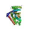
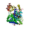
| Title | Crystal structure of Activin receptor type-IIA (ACVR2A) kinase domain in complex with dihydro-Bauerine C | ||||||
 Components Components | ACTIVIN RECEPTOR TYPE-2A | ||||||
 Keywords Keywords |  TRANSFERASE / PROTEIN KINASE TRANSFERASE / PROTEIN KINASE | ||||||
| Function / homology |  Function and homology information Function and homology informationRegulation of signaling by NODAL / inhibin-betaglycan-ActRII complex / inhibin binding / penile erection / positive regulation of activin receptor signaling pathway / activin receptor activity / Sertoli cell proliferation / sperm ejaculation / BMP receptor activity / embryonic skeletal system development ...Regulation of signaling by NODAL / inhibin-betaglycan-ActRII complex / inhibin binding / penile erection / positive regulation of activin receptor signaling pathway / activin receptor activity / Sertoli cell proliferation / sperm ejaculation / BMP receptor activity / embryonic skeletal system development / activin receptor complex / receptor protein serine/threonine kinase / transmembrane receptor protein serine/threonine kinase activity / Signaling by BMP / activin binding / cellular response to BMP stimulus / activin receptor signaling pathway / Signaling by Activin / Signaling by NODAL / gastrulation with mouth forming second / regulation of nitric oxide biosynthetic process / determination of left/right symmetry / anterior/posterior pattern specification / cell surface receptor protein serine/threonine kinase signaling pathway / odontogenesis of dentin-containing tooth / growth factor binding / mesoderm development / positive regulation of SMAD protein signal transduction / BMP signaling pathway / positive regulation of bone mineralization / positive regulation of osteoblast differentiation / coreceptor activity / positive regulation of erythrocyte differentiation / PDZ domain binding / cellular response to growth factor stimulus / : / spermatogenesis / receptor complex / positive regulation of protein phosphorylation / phosphorylation / protein serine/threonine kinase activity / cell surface / positive regulation of transcription by RNA polymerase II / ATP binding / metal ion binding / plasma membrane / cytoplasmSimilarity search - Function | ||||||
| Biological species |  HOMO SAPIENS (human) HOMO SAPIENS (human) | ||||||
| Method | X-RAY DIFFRACTION / SYNCHROTRON / MOLECULAR REPLACEMENT / Resolution: 2.05 Å | ||||||
 Authors Authors | Williams, E. / Chaikuad, A. / Canning, P. / Kochan, G. / Mahajan, P. / Cooper, C.D.O. / Beltrami, A. / Krojer, T. / Pohl, B. / Bracher, F. ...Williams, E. / Chaikuad, A. / Canning, P. / Kochan, G. / Mahajan, P. / Cooper, C.D.O. / Beltrami, A. / Krojer, T. / Pohl, B. / Bracher, F. / Arrowsmith, C.H. / Edwards, A.M. / Bountra, C. / von Delft, F. / Bullock, A. | ||||||
 Citation Citation | Journal: To be Published Title: Crystal Structure of Activin Receptor Type-Iia (Acvr2A) Kinase Domain in Complex with a Beta- Carboline Inhibitor Authors: Williams, E. / Chaikuad, A. / Canning, P. / Mahajan, P. / Cooper, C.D.O. / Beltrami, A. / Krojer, T. / Huber, K. / Bracher, F. / von Delft, F. / Arrowsmith, C.H. / Edwards, A.M. / Bountra, C. / Bullock, A. | ||||||
| History |
|
- Structure visualization
| Structure viewer | Molecule: MolmilJmol/JSmol |
|---|
- Downloads & links
-Download
| PDBx/mmCIF format | 4asx.cif.gz | 258.6 KB | Display | PDBx/mmCIF format |
|---|---|---|---|---|
| PDB format | pdb4asx.ent.gz | 209.9 KB | Display | PDB format |
| PDBx/mmJSON format | 4asx.json.gz | Tree view | PDBx/mmJSON format | |
| Others |  Other downloads Other downloads |
-Validation report
| Arichive directory | https://data.pdbj.org/pub/pdb/validation_reports/as/4asxftp://data.pdbj.org/pub/pdb/validation_reports/as/4asx | HTTPS FTP |
|---|
-Related structure data
| Related structure data |  3q4tS S: Starting model for refinement |
|---|---|
| Similar structure data |


-Links
 PDBj
PDBj

- Assembly
| Deposited unit | 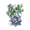
| ||||||||||||||||||||||||||||||||||||||||||||||||||||||||||||||||||||||||||||||||||||||||||||||||||||||||||||||||||||||||||||||||||||||||||||||
|---|---|---|---|---|---|---|---|---|---|---|---|---|---|---|---|---|---|---|---|---|---|---|---|---|---|---|---|---|---|---|---|---|---|---|---|---|---|---|---|---|---|---|---|---|---|---|---|---|---|---|---|---|---|---|---|---|---|---|---|---|---|---|---|---|---|---|---|---|---|---|---|---|---|---|---|---|---|---|---|---|---|---|---|---|---|---|---|---|---|---|---|---|---|---|---|---|---|---|---|---|---|---|---|---|---|---|---|---|---|---|---|---|---|---|---|---|---|---|---|---|---|---|---|---|---|---|---|---|---|---|---|---|---|---|---|---|---|---|---|---|---|---|---|
| 1 | 
| ||||||||||||||||||||||||||||||||||||||||||||||||||||||||||||||||||||||||||||||||||||||||||||||||||||||||||||||||||||||||||||||||||||||||||||||
| 2 | 
| ||||||||||||||||||||||||||||||||||||||||||||||||||||||||||||||||||||||||||||||||||||||||||||||||||||||||||||||||||||||||||||||||||||||||||||||
| Unit cell |
| ||||||||||||||||||||||||||||||||||||||||||||||||||||||||||||||||||||||||||||||||||||||||||||||||||||||||||||||||||||||||||||||||||||||||||||||
| Components on special symmetry positions |
| ||||||||||||||||||||||||||||||||||||||||||||||||||||||||||||||||||||||||||||||||||||||||||||||||||||||||||||||||||||||||||||||||||||||||||||||
| Noncrystallographic symmetry (NCS) | NCS domain:
NCS domain segments: Ens-ID: 1
NCS oper:
|
-Components
| #1: Protein | Mass: 36548.766 Da / Num. of mol.: 2 / Fragment: KINASE DOMAIN, RESIDUES 191-488 Source method: isolated from a genetically manipulated source Source: (gene. exp.) HOMO SAPIENS (human) / Plasmid: PFB-LIC-BSE / Cell line (production host): SF9 / Production host:   SPODOPTERA FRUGIPERDA (fall armyworm) SPODOPTERA FRUGIPERDA (fall armyworm)References: UniProt: P27037, receptor protein serine/threonine kinase#2: Chemical |   Mass: 269.127 Da / Num. of mol.: 2 / Source method: obtained synthetically / Formula: C12H10Cl2N2O Mass: 269.127 Da / Num. of mol.: 2 / Source method: obtained synthetically / Formula: C12H10Cl2N2O#3: Chemical | ChemComp-EDO / Ethylene glycol  Mass: 62.068 Da / Num. of mol.: 9 / Source method: obtained synthetically / Formula: C2H6O2 Mass: 62.068 Da / Num. of mol.: 9 / Source method: obtained synthetically / Formula: C2H6O2#4: Water | ChemComp-HOH / | Water Mass: 18.015 Da / Num. of mol.: 321 / Source method: isolated from a natural source / Formula: H2O Mass: 18.015 Da / Num. of mol.: 321 / Source method: isolated from a natural source / Formula: H2O |
|---|
-Experimental details
-Experiment
| Experiment | Method: X-RAY DIFFRACTION / Number of used crystals: 1 |
|---|
- Sample preparation
| Crystal | Density Matthews: 2.62 Å3/Da / Density % sol: 53.07 % / Description: NONE |
|---|---|
| Crystal grow | Details: 20 % PEG 3350 0.20 M NA(MALONATE) |
-Data collection
| Diffraction | Mean temperature: 100 K |
|---|---|
| Diffraction source | Source: SYNCHROTRON / Site: Diamond  / Beamline: I04 / Wavelength: 0.9611 / Beamline: I04 / Wavelength: 0.9611 |
| Detector | Type: ADSC CCD / Detector: CCD / Date: Jul 28, 2011 |
| Radiation | Protocol: SINGLE WAVELENGTH / Monochromatic (M) / Laue (L): M / Scattering type: x-ray |
| Radiation wavelength | Wavelength: 0.9611 Å / Relative weight: 1 |
| Reflection | Resolution: 2.05→19.99 Å / Num. obs: 47777 / % possible obs: 99.7 % / Observed criterion σ(I): 2 / Redundancy: 4.5 % / Biso Wilson estimate: 25.2 Å2 / Rmerge(I) obs: 0.13 / Net I/σ(I): 8 |
| Reflection shell | Resolution: 2.05→2.16 Å / Redundancy: 4.6 % / Rmerge(I) obs: 0.82 / Mean I/σ(I) obs: 1.9 / % possible all: 99.8 |
- Processing
| Software |
| ||||||||||||||||||||||||||||||||||||||||||||||||||||||||||||||||||||||||||||||||||||||||||||||||||||||||||||||||||||||||||||||||||||||||||||||||||||||||||||||||||||||||||||||||||||||
|---|---|---|---|---|---|---|---|---|---|---|---|---|---|---|---|---|---|---|---|---|---|---|---|---|---|---|---|---|---|---|---|---|---|---|---|---|---|---|---|---|---|---|---|---|---|---|---|---|---|---|---|---|---|---|---|---|---|---|---|---|---|---|---|---|---|---|---|---|---|---|---|---|---|---|---|---|---|---|---|---|---|---|---|---|---|---|---|---|---|---|---|---|---|---|---|---|---|---|---|---|---|---|---|---|---|---|---|---|---|---|---|---|---|---|---|---|---|---|---|---|---|---|---|---|---|---|---|---|---|---|---|---|---|---|---|---|---|---|---|---|---|---|---|---|---|---|---|---|---|---|---|---|---|---|---|---|---|---|---|---|---|---|---|---|---|---|---|---|---|---|---|---|---|---|---|---|---|---|---|---|---|---|---|
| Refinement | Method to determine structure: MOLECULAR REPLACEMENT Starting model: PDB ENTRY 3Q4T Resolution: 2.05→19.99 Å / Cor.coef. Fo:Fc: 0.951 / Cor.coef. Fo:Fc free: 0.937 / SU B: 7.666 / SU ML: 0.108 / Cross valid method: THROUGHOUT / ESU R: 0.184 / ESU R Free: 0.161 / Stereochemistry target values: MAXIMUM LIKELIHOOD Details: HYDROGENS HAVE BEEN ADDED IN THE RIDING POSITIONS. U VALUES WITH TLS ADDED
| ||||||||||||||||||||||||||||||||||||||||||||||||||||||||||||||||||||||||||||||||||||||||||||||||||||||||||||||||||||||||||||||||||||||||||||||||||||||||||||||||||||||||||||||||||||||
| Solvent computation | Ion probe radii: 0.8 Å / Shrinkage radii: 0.8 Å / VDW probe radii: 1.2 Å / Solvent model: MASK | ||||||||||||||||||||||||||||||||||||||||||||||||||||||||||||||||||||||||||||||||||||||||||||||||||||||||||||||||||||||||||||||||||||||||||||||||||||||||||||||||||||||||||||||||||||||
| Displacement parameters | Biso mean: 25.745 Å2
| ||||||||||||||||||||||||||||||||||||||||||||||||||||||||||||||||||||||||||||||||||||||||||||||||||||||||||||||||||||||||||||||||||||||||||||||||||||||||||||||||||||||||||||||||||||||
| Refinement step | Cycle: LAST / Resolution: 2.05→19.99 Å
| ||||||||||||||||||||||||||||||||||||||||||||||||||||||||||||||||||||||||||||||||||||||||||||||||||||||||||||||||||||||||||||||||||||||||||||||||||||||||||||||||||||||||||||||||||||||
| Refine LS restraints |
|