 Movie
Movie Controller

[English] 日本語
 Yorodumi
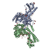
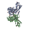
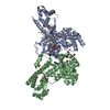
- PDB-6wk1: SETD3 in Complex with an Actin Peptide with His73 Replaced with M... -
+ Open data

- Basic information
| Entry | Database: PDB / ID: 6wk1 | ||||||
|---|---|---|---|---|---|---|---|
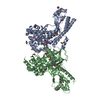
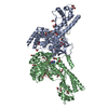
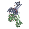
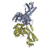
| Title | SETD3 in Complex with an Actin Peptide with His73 Replaced with Methionine | ||||||
 Components Components |
| ||||||
 Keywords Keywords |  TRANSFERASE / TRANSFERASE-STRUCTURAL PROTEIN complex TRANSFERASE / TRANSFERASE-STRUCTURAL PROTEIN complex | ||||||
| Function / homology |  Function and homology information Function and homology informationbasal body patch / protein-histidine N-methyltransferase / peptidyl-histidine methylation / regulation of uterine smooth muscle contraction / protein-L-histidine N-tele-methyltransferase activity / actin modification / tight junction assembly / regulation of transepithelial transport / structural constituent of postsynaptic actin cytoskeleton / profilin binding ...basal body patch / protein-histidine N-methyltransferase / peptidyl-histidine methylation / regulation of uterine smooth muscle contraction / protein-L-histidine N-tele-methyltransferase activity / actin modification / tight junction assembly / regulation of transepithelial transport / structural constituent of postsynaptic actin cytoskeleton / profilin binding / morphogenesis of a polarized epithelium / protein localization to bicellular tight junction / Formation of annular gap junctions / histone H3K36 methyltransferase activity / dense body / Gap junction degradation / Cell-extracellular matrix interactions / regulation of stress fiber assembly / Adherens junctions interactions / histone H3K4 methyltransferase activity / Sensory processing of sound by outer hair cells of the cochlea / Interaction between L1 and Ankyrins / Sensory processing of sound by inner hair cells of the cochlea / sarcomere organization / NuA4 histone acetyltransferase complex / regulation of synaptic vesicle endocytosis / apical junction complex / regulation of focal adhesion assembly / maintenance of blood-brain barrier / positive regulation of wound healing / myofibril / positive regulation of muscle cell differentiation / Recycling pathway of L1 / filamentous actin / calyx of Held / EPH-ephrin mediated repulsion of cells / RHO GTPases Activate WASPs and WAVEs / RHO GTPases activate IQGAPs / RHOBTB2 GTPase cycle / phagocytic vesicle / EPHB-mediated forward signaling / axonogenesis / cell motility / actin filament / Translocation of SLC2A4 (GLUT4) to the plasma membrane / RHO GTPases Activate Formins / FCGR3A-mediated phagocytosis / Hydrolases; Acting on acid anhydrides; Acting on acid anhydrides to facilitate cellular and subcellular movement / Signaling by high-kinase activity BRAF mutants / Schaffer collateral - CA1 synapse / MAP2K and MAPK activation / structural constituent of cytoskeleton / Regulation of actin dynamics for phagocytic cup formation / PKMTs methylate histone lysines / platelet aggregation / VEGFA-VEGFR2 Pathway / cellular response to type II interferon / Signaling by RAF1 mutants / Signaling by moderate kinase activity BRAF mutants / Paradoxical activation of RAF signaling by kinase inactive BRAF / Signaling downstream of RAS mutants / Signaling by BRAF and RAF1 fusions / cell-cell junction / Clathrin-mediated endocytosis / actin binding / angiogenesis / blood microparticle / RNA polymerase II-specific DNA-binding transcription factor binding / transcription coactivator activity / cytoskeleton / hydrolase activity / positive regulation of cell migration / axon / focal adhesion / synapse / ubiquitin protein ligase binding / chromatin / positive regulation of gene expression / protein kinase binding / positive regulation of DNA-templated transcription / positive regulation of transcription by RNA polymerase II / extracellular space / extracellular exosome / nucleoplasm / ATP binding / membrane / identical protein binding / nucleus / plasma membrane / cytosol / cytoplasmSimilarity search - Function | ||||||
| Biological species |  Homo sapiens (human) Homo sapiens (human) | ||||||
| Method | X-RAY DIFFRACTION / SYNCHROTRON / MOLECULAR REPLACEMENT / Resolution: 1.89 Å | ||||||
 Authors Authors | Dai, S. / Horton, J.R. / Cheng, X. | ||||||
| Funding support |  United States, 1items United States, 1items
| ||||||
 Citation Citation | Journal: J.Biol.Chem. / Year: 2020 Title: Characterization of SETD3 methyltransferase-mediated protein methionine methylation. Authors: Dai, S. / Holt, M.V. / Horton, J.R. / Woodcock, C.B. / Patel, A. / Zhang, X. / Young, N.L. / Wilkinson, A.W. / Cheng, X. | ||||||
| History |
|
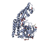
- Structure visualization
| Structure viewer | Molecule: MolmilJmol/JSmol |
|---|
- Downloads & links
-Download
| PDBx/mmCIF format | 6wk1.cif.gz | 293.6 KB | Display | PDBx/mmCIF format |
|---|---|---|---|---|
| PDB format | pdb6wk1.ent.gz | 186.3 KB | Display | PDB format |
| PDBx/mmJSON format | 6wk1.json.gz | Tree view | PDBx/mmJSON format | |
| Others |  Other downloads Other downloads |
-Validation report
| Arichive directory | https://data.pdbj.org/pub/pdb/validation_reports/wk/6wk1ftp://data.pdbj.org/pub/pdb/validation_reports/wk/6wk1 | HTTPS FTP |
|---|
-Related structure data
| Related structure data |  6wk2C  6mbjS S: Starting model for refinement C: citing same article ( |
|---|---|
| Similar structure data |

-Links
 PDBj
PDBj


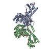
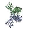
- Assembly
| Deposited unit | 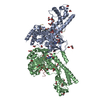
| ||||||||||||
|---|---|---|---|---|---|---|---|---|---|---|---|---|---|
| 1 |
| ||||||||||||
| 2 | 
| ||||||||||||
| 3 | 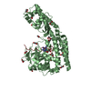
| ||||||||||||
| Unit cell |
|
-Components
-Protein/peptide / Protein , 2 types, 4 molecules YZAB
| #1: Protein/peptide | / Gamma-actin Mass: 2861.296 Da / Num. of mol.: 2 / Mutation: H73M / Source method: obtained synthetically / Source: (synth.) Homo sapiens (human) / References: UniProt: P63261#2: Protein | Mass: 67342.047 Da / Num. of mol.: 2 Source method: isolated from a genetically manipulated source Source: (gene. exp.) Homo sapiens (human) / Gene: SETD3, C14orf154 / Production host:  Escherichia coli (E. coli) Escherichia coli (E. coli)References: UniProt: Q86TU7, protein-histidine N-methyltransferase |
|---|
-Non-polymers , 5 types, 656 molecules 


| #3: Chemical | ChemComp-GOL / Glycerol Mass: 92.094 Da / Num. of mol.: 6 / Source method: obtained synthetically / Formula: C3H8O3 Mass: 92.094 Da / Num. of mol.: 6 / Source method: obtained synthetically / Formula: C3H8O3#4: Chemical | ChemComp-EDO / Ethylene glycol Mass: 62.068 Da / Num. of mol.: 46 / Source method: obtained synthetically / Formula: C2H6O2 Mass: 62.068 Da / Num. of mol.: 46 / Source method: obtained synthetically / Formula: C2H6O2#5: Chemical | S-Adenosyl-L-homocysteine Type: L-peptide linking / Mass: 384.411 Da / Num. of mol.: 2 / Source method: obtained synthetically / Formula: C14H20N6O5S Type: L-peptide linking / Mass: 384.411 Da / Num. of mol.: 2 / Source method: obtained synthetically / Formula: C14H20N6O5S#6: Chemical | ChemComp-ACT / | Acetate Mass: 59.044 Da / Num. of mol.: 1 / Source method: obtained synthetically / Formula: C2H3O2 Mass: 59.044 Da / Num. of mol.: 1 / Source method: obtained synthetically / Formula: C2H3O2#7: Water | ChemComp-HOH / | WaterMass: 18.015 Da / Num. of mol.: 601 / Source method: isolated from a natural source / Formula: H2O |
|---|
-Details
| Has ligand of interest | N |
|---|
-Experimental details
-Experiment
| Experiment | Method: X-RAY DIFFRACTION / Number of used crystals: 1 |
|---|
- Sample preparation
| Crystal | Density Matthews: 2.51 Å3/Da / Density % sol: 51.07 % |
|---|---|
| Crystal grow | Temperature: 292 K / Method: vapor diffusion, sitting drop / pH: 5.6 Details: 0.2 M ammonium acetate, 0.1 M sodium citrate tribasic dihydrate pH 5.6 and 30% (w/v) polyethylene glycol 4000 |
-Data collection
| Diffraction | Mean temperature: 100 K / Serial crystal experiment: N |
|---|---|
| Diffraction source | Source: SYNCHROTRON / Site: APS / Beamline: 22-ID / Wavelength: 1 Å |
| Detector | Type: DECTRIS EIGER X 16M / Detector: PIXEL / Date: Dec 13, 2019 |
| Radiation | Protocol: SINGLE WAVELENGTH / Monochromatic (M) / Laue (L): M / Scattering type: x-ray |
| Radiation wavelength | Wavelength: 1 Å / Relative weight: 1 |
| Reflection | Resolution: 1.89→36 Å / Num. obs: 102262 / % possible obs: 93.6 % / Redundancy: 4.7 % / Biso Wilson estimate: 23.23 Å2 / CC1/2: 0.986 / Net I/σ(I): 10.2 |
| Reflection shell | Resolution: 1.89→1.96 Å / Num. unique obs: 8146 / CC1/2: 0.526 |
- Processing
| Software |
| |||||||||||||||||||||||||||||||||||||||||||||||||||||||||||||||||||||||||||||||||||||||||||||||||||||||||
|---|---|---|---|---|---|---|---|---|---|---|---|---|---|---|---|---|---|---|---|---|---|---|---|---|---|---|---|---|---|---|---|---|---|---|---|---|---|---|---|---|---|---|---|---|---|---|---|---|---|---|---|---|---|---|---|---|---|---|---|---|---|---|---|---|---|---|---|---|---|---|---|---|---|---|---|---|---|---|---|---|---|---|---|---|---|---|---|---|---|---|---|---|---|---|---|---|---|---|---|---|---|---|---|---|---|---|
| Refinement | Method to determine structure: MOLECULAR REPLACEMENT Starting model: 6MBJ Resolution: 1.89→36 Å / SU ML: 0.2308 / Cross valid method: FREE R-VALUE / σ(F): 1.35 / Phase error: 25.1001 / Stereochemistry target values: GeoStd + Monomer Library
| |||||||||||||||||||||||||||||||||||||||||||||||||||||||||||||||||||||||||||||||||||||||||||||||||||||||||
| Solvent computation | Shrinkage radii: 0.9 Å / VDW probe radii: 1.11 Å / Solvent model: FLAT BULK SOLVENT MODEL | |||||||||||||||||||||||||||||||||||||||||||||||||||||||||||||||||||||||||||||||||||||||||||||||||||||||||
| Displacement parameters | Biso mean: 28.19 Å2 | |||||||||||||||||||||||||||||||||||||||||||||||||||||||||||||||||||||||||||||||||||||||||||||||||||||||||
| Refinement step | Cycle: LAST / Resolution: 1.89→36 Å
| |||||||||||||||||||||||||||||||||||||||||||||||||||||||||||||||||||||||||||||||||||||||||||||||||||||||||
| Refine LS restraints |
| |||||||||||||||||||||||||||||||||||||||||||||||||||||||||||||||||||||||||||||||||||||||||||||||||||||||||
| LS refinement shell |
|
