 Movie
Movie Controller

[English] 日本語
 Yorodumi
- PDB-6wid: Nucleotide incorporation intermediate into quaternary complex of ... -
+ Open data

- Basic information
| Entry | Database: PDB / ID: 6wid | |||||||||
|---|---|---|---|---|---|---|---|---|---|---|
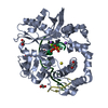
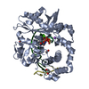
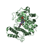
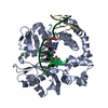
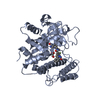
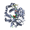
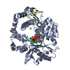
| Title | Nucleotide incorporation intermediate into quaternary complex of human Polymerase Mu on a complementary DNA double-strand break substrate | |||||||||
 Components Components |
| |||||||||
 Keywords Keywords | TRANSFERASE/DNA / Family X polymerase / nonhomologous end-joining / DNA double-strand break repair / TRANSFERASE-DNA complex | |||||||||
| Function / homology |  Function and homology information Function and homology informationNonhomologous End-Joining (NHEJ) / double-strand break repair via nonhomologous end joining / DNA recombination /  DNA-directed DNA polymerase / DNA-directed DNA polymerase activity / DNA binding / nucleoplasm / metal ion binding / nucleus DNA-directed DNA polymerase / DNA-directed DNA polymerase activity / DNA binding / nucleoplasm / metal ion binding / nucleusSimilarity search - Function | |||||||||
| Biological species |  Homo sapiens (human) Homo sapiens (human)synthetic construct (others) | |||||||||
| Method | X-RAY DIFFRACTION / SYNCHROTRON / MOLECULAR REPLACEMENT / Resolution: 1.5 Å | |||||||||
 Authors Authors | Kaminski, A.M. / Kunkel, T.A. / Pedersen, L.C. / Bebenek, K. | |||||||||
| Funding support |  United States, 2items United States, 2items
| |||||||||
 Citation Citation | Journal: Nat Commun / Year: 2020 Title: Structural snapshots of human DNA polymerase mu engaged on a DNA double-strand break. Authors: Kaminski, A.M. / Pryor, J.M. / Ramsden, D.A. / Kunkel, T.A. / Pedersen, L.C. / Bebenek, K. | |||||||||
| History |
|
- Structure visualization
| Structure viewer | Molecule: MolmilJmol/JSmol |
|---|
- Downloads & links
-Download
| PDBx/mmCIF format | 6wid.cif.gz | 210.7 KB | Display | PDBx/mmCIF format |
|---|---|---|---|---|
| PDB format | pdb6wid.ent.gz | 141.6 KB | Display | PDB format |
| PDBx/mmJSON format | 6wid.json.gz | Tree view | PDBx/mmJSON format | |
| Others |  Other downloads Other downloads |
-Validation report
| Arichive directory | https://data.pdbj.org/pub/pdb/validation_reports/wi/6widftp://data.pdbj.org/pub/pdb/validation_reports/wi/6wid | HTTPS FTP |
|---|
-Related structure data
| Related structure data |  6wicC  6wieC  4m04S S: Starting model for refinement C: citing same article ( |
|---|---|
| Similar structure data |


-Links
 PDBj
PDBj


- Assembly
| Deposited unit | 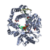
| ||||||||||||
|---|---|---|---|---|---|---|---|---|---|---|---|---|---|
| 1 |
| ||||||||||||
| Unit cell |
|
-Components
-Protein , 1 types, 1 molecules A
| #1: Protein | Mass: 40054.434 Da / Num. of mol.: 1 / Fragment: UNP residues 132-494 / Mutation: P398-P410 deletion replaced by G410 linker Source method: isolated from a genetically manipulated source Source: (gene. exp.) Homo sapiens (human) / Gene: POLM, polmu / Plasmid: pGEXM / Production host:  Escherichia coli BL21(DE3) (bacteria) / Strain (production host): Rosetta2 (DE3) / References: UniProt: Q9NP87, DNA-directed DNA polymerase Escherichia coli BL21(DE3) (bacteria) / Strain (production host): Rosetta2 (DE3) / References: UniProt: Q9NP87, DNA-directed DNA polymerase |
|---|
-DNA chain , 4 types, 4 molecules TUPD
| #2: DNA chain | Mass: 1809.217 Da / Num. of mol.: 1 / Source method: obtained synthetically / Source: (synth.) synthetic construct (others) |
|---|---|
| #3: DNA chain | Mass: 886.637 Da / Num. of mol.: 1 / Source method: obtained synthetically / Source: (synth.) synthetic construct (others) |
| #4: DNA chain | Mass: 1495.023 Da / Num. of mol.: 1 / Source method: obtained synthetically / Source: (synth.) synthetic construct (others) |
| #5: DNA chain | Mass: 1191.818 Da / Num. of mol.: 1 / Source method: obtained synthetically / Source: (synth.) synthetic construct (others) |
-Non-polymers , 9 types, 360 molecules 


| #6: Chemical | ChemComp-DUP /  Mass: 467.157 Da / Num. of mol.: 1 / Source method: obtained synthetically / Formula: C9H16N3O13P3 / Feature type: SUBJECT OF INVESTIGATION Mass: 467.157 Da / Num. of mol.: 1 / Source method: obtained synthetically / Formula: C9H16N3O13P3 / Feature type: SUBJECT OF INVESTIGATION | ||||||||||||
|---|---|---|---|---|---|---|---|---|---|---|---|---|---|
| #7: Chemical | ChemComp-PPV / Pyrophosphate Mass: 177.975 Da / Num. of mol.: 1 / Source method: obtained synthetically / Formula: H4O7P2 / Feature type: SUBJECT OF INVESTIGATION Mass: 177.975 Da / Num. of mol.: 1 / Source method: obtained synthetically / Formula: H4O7P2 / Feature type: SUBJECT OF INVESTIGATION | ||||||||||||
| #8: Chemical |  Mass: 24.305 Da / Num. of mol.: 2 / Source method: obtained synthetically / Formula: Mg / Feature type: SUBJECT OF INVESTIGATION Mass: 24.305 Da / Num. of mol.: 2 / Source method: obtained synthetically / Formula: Mg / Feature type: SUBJECT OF INVESTIGATION#9: Chemical | ChemComp-NA / |  Mass: 22.990 Da / Num. of mol.: 1 / Source method: obtained synthetically / Formula: Na Mass: 22.990 Da / Num. of mol.: 1 / Source method: obtained synthetically / Formula: Na#10: Chemical | ChemComp-CL / | Chloride Mass: 35.453 Da / Num. of mol.: 1 / Source method: obtained synthetically / Formula: Cl Mass: 35.453 Da / Num. of mol.: 1 / Source method: obtained synthetically / Formula: Cl#11: Chemical | ChemComp-1PE / | Polyethylene glycol Mass: 238.278 Da / Num. of mol.: 1 / Source method: obtained synthetically / Formula: C10H22O6 / Comment: precipitant*YM Mass: 238.278 Da / Num. of mol.: 1 / Source method: obtained synthetically / Formula: C10H22O6 / Comment: precipitant*YM#12: Chemical | Glycerol Mass: 92.094 Da / Num. of mol.: 2 / Source method: obtained synthetically / Formula: C3H8O3 Mass: 92.094 Da / Num. of mol.: 2 / Source method: obtained synthetically / Formula: C3H8O3#13: Chemical | ChemComp-PEG / | Diethylene glycol Mass: 106.120 Da / Num. of mol.: 1 / Source method: obtained synthetically / Formula: C4H10O3 Mass: 106.120 Da / Num. of mol.: 1 / Source method: obtained synthetically / Formula: C4H10O3#14: Water | ChemComp-HOH / | WaterMass: 18.015 Da / Num. of mol.: 350 / Source method: isolated from a natural source / Formula: H2O |
-Details
| Has ligand of interest | Y |
|---|
-Experimental details
-Experiment
| Experiment | Method: X-RAY DIFFRACTION / Number of used crystals: 1 |
|---|
- Sample preparation
| Crystal | Density Matthews: 2.43 Å3/Da / Density % sol: 49.4 % |
|---|---|
| Crystal grow | Temperature: 277 K / Method: vapor diffusion, sitting drop / pH: 5.6 Details: 40-45.5 mM MES, pH 5.6, 0.16-0.182 M potassium chloride, 8.2-9.1 mM magnesium sulfate, 8.2-9.1% w/v PEG400 |
-Data collection
| Diffraction | Mean temperature: 93 K / Serial crystal experiment: N |
|---|---|
| Diffraction source | Source: SYNCHROTRON / Site: APS / Beamline: 22-ID / Wavelength: 1 Å |
| Detector | Type: DECTRIS EIGER X 16M / Detector: PIXEL / Date: Dec 5, 2019 |
| Radiation | Monochromator: double crystal Si(111) / Protocol: SINGLE WAVELENGTH / Monochromatic (M) / Laue (L): M / Scattering type: x-ray |
| Radiation wavelength | Wavelength: 1 Å / Relative weight: 1 |
| Reflection | Resolution: 1.5→50 Å / Num. obs: 71251 / % possible obs: 99.8 % / Redundancy: 6 % / Biso Wilson estimate: 14.06 Å2 / CC1/2: 0.998 / CC star: 0.999 / Rpim(I) all: 0.037 / Rrim(I) all: 0.091 / Rsym value: 0.083 / Χ2: 1.215 / Net I/σ(I): 28.5 |
| Reflection shell | Resolution: 1.5→1.53 Å / Redundancy: 5.5 % / Mean I/σ(I) obs: 2.24 / Num. unique obs: 3524 / CC1/2: 0.848 / CC star: 0.958 / Rpim(I) all: 0.255 / Rrim(I) all: 0.606 / Rsym value: 0.548 / Χ2: 0.42 / % possible all: 99.9 |
- Processing
| Software |
| |||||||||||||||||||||||||||||||||||||||||||||||||||||||||||||||||||||||||||||||||||||||||||||||||||||||||||||||||||||||||||||||||||||||||||||||||||||||||||||||||||||||||||||||
|---|---|---|---|---|---|---|---|---|---|---|---|---|---|---|---|---|---|---|---|---|---|---|---|---|---|---|---|---|---|---|---|---|---|---|---|---|---|---|---|---|---|---|---|---|---|---|---|---|---|---|---|---|---|---|---|---|---|---|---|---|---|---|---|---|---|---|---|---|---|---|---|---|---|---|---|---|---|---|---|---|---|---|---|---|---|---|---|---|---|---|---|---|---|---|---|---|---|---|---|---|---|---|---|---|---|---|---|---|---|---|---|---|---|---|---|---|---|---|---|---|---|---|---|---|---|---|---|---|---|---|---|---|---|---|---|---|---|---|---|---|---|---|---|---|---|---|---|---|---|---|---|---|---|---|---|---|---|---|---|---|---|---|---|---|---|---|---|---|---|---|---|---|---|---|---|---|
| Refinement | Method to determine structure: MOLECULAR REPLACEMENT Starting model: PDB entry 4M04 Resolution: 1.5→31.02 Å / SU ML: 0.1371 / Cross valid method: FREE R-VALUE / σ(F): 1.37 / Phase error: 15.8136
| |||||||||||||||||||||||||||||||||||||||||||||||||||||||||||||||||||||||||||||||||||||||||||||||||||||||||||||||||||||||||||||||||||||||||||||||||||||||||||||||||||||||||||||||
| Solvent computation | Shrinkage radii: 0.9 Å / VDW probe radii: 1.11 Å | |||||||||||||||||||||||||||||||||||||||||||||||||||||||||||||||||||||||||||||||||||||||||||||||||||||||||||||||||||||||||||||||||||||||||||||||||||||||||||||||||||||||||||||||
| Displacement parameters | Biso mean: 19.95 Å2 | |||||||||||||||||||||||||||||||||||||||||||||||||||||||||||||||||||||||||||||||||||||||||||||||||||||||||||||||||||||||||||||||||||||||||||||||||||||||||||||||||||||||||||||||
| Refinement step | Cycle: LAST / Resolution: 1.5→31.02 Å
| |||||||||||||||||||||||||||||||||||||||||||||||||||||||||||||||||||||||||||||||||||||||||||||||||||||||||||||||||||||||||||||||||||||||||||||||||||||||||||||||||||||||||||||||
| Refine LS restraints |
| |||||||||||||||||||||||||||||||||||||||||||||||||||||||||||||||||||||||||||||||||||||||||||||||||||||||||||||||||||||||||||||||||||||||||||||||||||||||||||||||||||||||||||||||
| LS refinement shell |
| |||||||||||||||||||||||||||||||||||||||||||||||||||||||||||||||||||||||||||||||||||||||||||||||||||||||||||||||||||||||||||||||||||||||||||||||||||||||||||||||||||||||||||||||
| Refinement TLS params. | Method: refined / Refine-ID: X-RAY DIFFRACTION
| |||||||||||||||||||||||||||||||||||||||||||||||||||||||||||||||||||||||||||||||||||||||||||||||||||||||||||||||||||||||||||||||||||||||||||||||||||||||||||||||||||||||||||||||
| Refinement TLS group |
|
