 Movie
Movie Controller


+ Open data

- Basic information
| Entry | Database: PDB / ID: 6wib | ||||||
|---|---|---|---|---|---|---|---|
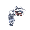
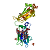
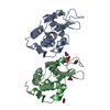
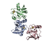
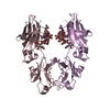
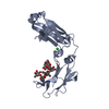
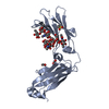
| Title | Next generation monomeric IgG4 Fc | ||||||
 Components Components | Immunoglobulin heavy constant gamma 4 | ||||||
 Keywords Keywords |  IMMUNE SYSTEM / antibody constant region / fragment crystallizable / mutated IMMUNE SYSTEM / antibody constant region / fragment crystallizable / mutated | ||||||
| Function / homology |  Function and homology information Function and homology informationClassical antibody-mediated complement activation / IgG immunoglobulin complex / Initial triggering of complement / FCGR activation / Role of phospholipids in phagocytosis / immunoglobulin complex, circulating / immunoglobulin receptor binding / complement activation, classical pathway / FCGR3A-mediated IL10 synthesis / antigen binding ...Classical antibody-mediated complement activation / IgG immunoglobulin complex / Initial triggering of complement / FCGR activation / Role of phospholipids in phagocytosis / immunoglobulin complex, circulating / immunoglobulin receptor binding / complement activation, classical pathway / FCGR3A-mediated IL10 synthesis / antigen binding / Regulation of Complement cascade / FCGR3A-mediated phagocytosis / B cell receptor signaling pathway / Regulation of actin dynamics for phagocytic cup formation / antibacterial humoral response / Interleukin-4 and Interleukin-13 signaling / adaptive immune response / blood microparticle / extracellular space / extracellular exosome / extracellular region / plasma membraneSimilarity search - Function | ||||||
| Biological species |  Homo sapiens (human) Homo sapiens (human) | ||||||
| Method | X-RAY DIFFRACTION / SYNCHROTRON / MOLECULAR REPLACEMENT / Resolution: 2.55 Å | ||||||
 Authors Authors | Oganesyan, V.Y. / Shan, L. / Dall'Acqua, W. / van Dyk, N. | ||||||
 Citation Citation | Journal: Commun Biol / Year: 2021 Title: In vivo pharmacokinetic enhancement of monomeric Fc and monovalent bispecific designs through structural guidance. Authors: Shan, L. / Dyk, N.V. / Haskins, N. / Cook, K.M. / Rosenthal, K.L. / Mazor, R. / Dragulin-Otto, S. / Jiang, Y. / Wu, H. / Dall'Acqua, W.F. / Borrok, M.J. / Damschroder, M.M. / Oganesyan, V. | ||||||
| History |
|
- Structure visualization
| Structure viewer | Molecule: MolmilJmol/JSmol |
|---|
- Downloads & links
-Download
| PDBx/mmCIF format | 6wib.cif.gz | 62.8 KB | Display | PDBx/mmCIF format |
|---|---|---|---|---|
| PDB format | pdb6wib.ent.gz | 43 KB | Display | PDB format |
| PDBx/mmJSON format | 6wib.json.gz | Tree view | PDBx/mmJSON format | |
| Others |  Other downloads Other downloads |
-Validation report
| Arichive directory | https://data.pdbj.org/pub/pdb/validation_reports/wi/6wibftp://data.pdbj.org/pub/pdb/validation_reports/wi/6wib | HTTPS FTP |
|---|
-Related structure data
| Related structure data |  6wmhC 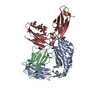 6wnaC  6wolC  5hvwS S: Starting model for refinement C: citing same article ( |
|---|---|
| Similar structure data |


-Links
 PDBj
PDBj


- Assembly
| Deposited unit | 
| ||||||||
|---|---|---|---|---|---|---|---|---|---|
| 1 |
| ||||||||
| Unit cell |
|
-Components
| #1: Protein | Mass: 23847.955 Da / Num. of mol.: 1 Mutation: L351F, T366R, P395K, F405R, Y407e, L432C, H433S, N434W, Y436L, T437C, Q438 deleted Source method: isolated from a genetically manipulated source Details: uniprot / Source: (gene. exp.) Homo sapiens (human) / Gene: IGHG4 / Production host:   Cricetulus griseus (Chinese hamster) / References: UniProt: P01861 Cricetulus griseus (Chinese hamster) / References: UniProt: P01861 | ||||
|---|---|---|---|---|---|
| #2: Polysaccharide | 2-acetamido-2-deoxy-beta-D-glucopyranose-(1-2)-alpha-D-mannopyranose-(1-3)-[2-acetamido-2-deoxy- ...2-acetamido-2-deoxy-beta-D-glucopyranose-(1-2)-alpha-D-mannopyranose-(1-3)-[2-acetamido-2-deoxy-beta-D-glucopyranose-(1-2)-alpha-D-mannopyranose-(1-6)]beta-D-mannopyranose-(1-4)-2-acetamido-2-deoxy-beta-D-glucopyranose-(1-4)-[alpha-L-fucopyranose-(1-6)]2-acetamido-2-deoxy-beta-D-glucopyranose / Mass: 1463.349 Da / Num. of mol.: 1 Source method: isolated from a genetically manipulated source | ||||
| #3: Chemical |   Mass: 65.409 Da / Num. of mol.: 3 / Source method: obtained synthetically / Formula: Zn Mass: 65.409 Da / Num. of mol.: 3 / Source method: obtained synthetically / Formula: Zn#4: Water | ChemComp-HOH / | Water Mass: 18.015 Da / Num. of mol.: 14 / Source method: isolated from a natural source / Formula: H2O Mass: 18.015 Da / Num. of mol.: 14 / Source method: isolated from a natural source / Formula: H2OHas ligand of interest | Y | |
-Experimental details
-Experiment
| Experiment | Method: X-RAY DIFFRACTION / Number of used crystals: 1 |
|---|
- Sample preparation
| Crystal | Density Matthews: 3.73 Å3/Da / Density % sol: 67.02 % |
|---|---|
| Crystal grow | Temperature: 298 K / Method: vapor diffusion / pH: 6.5 Details: 0.01 M zinc sulfate heptahydrate; 0.1 M morpholineethanesulfonic acid (MES) monohydrate, pH 6.5, and 25% (w/v) PEG 550 MME at a protein concentration of 5.5 mg/mL |
-Data collection
| Diffraction | Mean temperature: 100 K / Serial crystal experiment: N |
|---|---|
| Diffraction source | Source: SYNCHROTRON / Site: SSRL  / Beamline: BL9-2 / Wavelength: 1.19499 Å / Beamline: BL9-2 / Wavelength: 1.19499 Å |
| Detector | Type: DECTRIS PILATUS3 2M / Detector: PIXEL / Date: Aug 25, 2017 |
| Radiation | Protocol: SINGLE WAVELENGTH / Monochromatic (M) / Laue (L): M / Scattering type: x-ray |
| Radiation wavelength | Wavelength: 1.19499 Å / Relative weight: 1 |
| Reflection | Resolution: 2.55→35.92 Å / Num. obs: 11992 / % possible obs: 99.9 % / Redundancy: 7.3 % / CC1/2: 0.995 / Rmerge(I) obs: 0.154 / Rpim(I) all: 0.061 / Net I/σ(I): 11.8 |
| Reflection shell | Resolution: 2.55→2.66 Å / Rmerge(I) obs: 0.958 / Mean I/σ(I) obs: 2.3 / Num. unique obs: 1429 / CC1/2: 0.8 / Rpim(I) all: 0.377 |
- Processing
| Software |
| ||||||||||||||||||||||||||||||||||||||||||||||||||||||||||||
|---|---|---|---|---|---|---|---|---|---|---|---|---|---|---|---|---|---|---|---|---|---|---|---|---|---|---|---|---|---|---|---|---|---|---|---|---|---|---|---|---|---|---|---|---|---|---|---|---|---|---|---|---|---|---|---|---|---|---|---|---|---|
| Refinement | Method to determine structure: MOLECULAR REPLACEMENT Starting model: 5hvw Resolution: 2.55→35.92 Å / Cor.coef. Fo:Fc: 0.926 / Cor.coef. Fo:Fc free: 0.894 / SU B: 9.813 / SU ML: 0.206 / Cross valid method: THROUGHOUT / σ(F): 0 / ESU R: 0.366 / ESU R Free: 0.273 Details: HYDROGENS HAVE BEEN ADDED IN THE RIDING POSITIONS U VALUES : REFINED INDIVIDUALLY
| ||||||||||||||||||||||||||||||||||||||||||||||||||||||||||||
| Solvent computation | Ion probe radii: 0.8 Å / Shrinkage radii: 0.8 Å / VDW probe radii: 1.2 Å | ||||||||||||||||||||||||||||||||||||||||||||||||||||||||||||
| Displacement parameters | Biso max: 105.53 Å2 / Biso mean: 35.635 Å2 / Biso min: 10.49 Å2
| ||||||||||||||||||||||||||||||||||||||||||||||||||||||||||||
| Refinement step | Cycle: final / Resolution: 2.55→35.92 Å
| ||||||||||||||||||||||||||||||||||||||||||||||||||||||||||||
| Refine LS restraints |
| ||||||||||||||||||||||||||||||||||||||||||||||||||||||||||||
| LS refinement shell | Resolution: 2.55→2.613 Å / Rfactor Rfree error: 0
|
