 Movie
Movie Controller

[English] 日本語
 Yorodumi
- PDB-6w0l: Henipavirus W protein interacts with 14-3-3 to modulate host gene... -
+ Open data

- Basic information
| Entry | Database: PDB / ID: 6w0l | ||||||
|---|---|---|---|---|---|---|---|
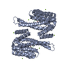
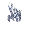
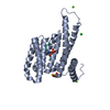
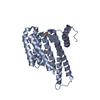
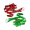
| Title | Henipavirus W protein interacts with 14-3-3 to modulate host gene expression | ||||||
 Components Components |
| ||||||
 Keywords Keywords |  PROTEIN BINDING / Complex / phosphorylation / nuclear import / nuclear localization PROTEIN BINDING / Complex / phosphorylation / nuclear import / nuclear localization | ||||||
| Function / homology |  Function and homology information Function and homology informationregulation of epidermal cell division / protein kinase C inhibitor activity / positive regulation of epidermal cell differentiation / keratinocyte development / keratinization / Regulation of localization of FOXO transcription factors / keratinocyte proliferation / phosphoserine residue binding / Activation of BAD and translocation to mitochondria / negative regulation of keratinocyte proliferation ...regulation of epidermal cell division / protein kinase C inhibitor activity / positive regulation of epidermal cell differentiation / keratinocyte development / keratinization / Regulation of localization of FOXO transcription factors / keratinocyte proliferation / phosphoserine residue binding / Activation of BAD and translocation to mitochondria / negative regulation of keratinocyte proliferation / establishment of skin barrier / SARS-CoV-2 targets host intracellular signalling and regulatory pathways / Chk1/Chk2(Cds1) mediated inactivation of Cyclin B:Cdk1 complex / negative regulation of stem cell proliferation / SARS-CoV-1 targets host intracellular signalling and regulatory pathways / RHO GTPases activate PKNs / protein kinase A signaling / protein export from nucleus / negative regulation of innate immune response / protein sequestering activity / TP53 Regulates Transcription of Genes Involved in G2 Cell Cycle Arrest / release of cytochrome c from mitochondria / positive regulation of protein export from nucleus / stem cell proliferation / Translocation of SLC2A4 (GLUT4) to the plasma membrane / TP53 Regulates Metabolic Genes / negative regulation of protein kinase activity / negative regulation of cysteine-type endopeptidase activity involved in apoptotic process / intrinsic apoptotic signaling pathway in response to DNA damage / positive regulation of cell growth / amyloid fibril formation / regulation of cell cycle / cadherin binding / virus-mediated perturbation of host defense response / host cell nucleus / protein kinase binding / negative regulation of transcription by RNA polymerase II / signal transduction / extracellular space / extracellular exosome / identical protein binding / nucleus / cytosol / cytoplasmSimilarity search - Function | ||||||
| Biological species |  Homo sapiens (human) Homo sapiens (human) Pteropus alecto (black flying fox) Pteropus alecto (black flying fox) | ||||||
| Method | X-RAY DIFFRACTION / SYNCHROTRON / MOLECULAR REPLACEMENT / molecular replacement / Resolution: 2.3 Å | ||||||
 Authors Authors | Edwards, M. / Hoad, M. / Tsimbalyuk, S. / Menicucci, A. / Messaoudi, I. / Forwood, J. / Basler, C. | ||||||
 Citation Citation | Journal: J.Virol. / Year: 2020 Title: Henipavirus W Proteins Interact with 14-3-3 To Modulate Host Gene Expression. Authors: Edwards, M.R. / Hoad, M. / Tsimbalyuk, S. / Menicucci, A.R. / Messaoudi, I. / Forwood, J.K. / Basler, C.F. | ||||||
| History |
|
- Structure visualization
| Structure viewer | Molecule: MolmilJmol/JSmol |
|---|
- Downloads & links
-Download
| PDBx/mmCIF format | 6w0l.cif.gz | 63.6 KB | Display | PDBx/mmCIF format |
|---|---|---|---|---|
| PDB format | pdb6w0l.ent.gz | 44.3 KB | Display | PDB format |
| PDBx/mmJSON format | 6w0l.json.gz | Tree view | PDBx/mmJSON format | |
| Others |  Other downloads Other downloads |
-Validation report
| Arichive directory | https://data.pdbj.org/pub/pdb/validation_reports/w0/6w0lftp://data.pdbj.org/pub/pdb/validation_reports/w0/6w0l | HTTPS FTP |
|---|
-Related structure data
| Related structure data |  4datS S: Starting model for refinement |
|---|---|
| Similar structure data |


-Links
 PDBj
PDBj


- Assembly
| Deposited unit | 
| ||||||||
|---|---|---|---|---|---|---|---|---|---|
| 1 | 
| ||||||||
| Unit cell |
| ||||||||
| Components on special symmetry positions |
|
-Components
| #1: Protein | Mass: 26542.914 Da / Num. of mol.: 1 Source method: isolated from a genetically manipulated source Source: (gene. exp.) Homo sapiens (human) / Gene: SFN, HME1 / Production host:  Escherichia coli (E. coli) / References: UniProt: P31947 Escherichia coli (E. coli) / References: UniProt: P31947 |
|---|---|
| #2: Protein/peptide | Mass: 1290.434 Da / Num. of mol.: 1 Source method: isolated from a genetically manipulated source Source: (gene. exp.) Pteropus alecto (black flying fox) / Production host: Escherichia coli (E. coli) / References: UniProt: P0C1C7*PLUS |
| #3: Chemical | ChemComp-CA /   Mass: 40.078 Da / Num. of mol.: 1 / Source method: obtained synthetically / Formula: Ca Mass: 40.078 Da / Num. of mol.: 1 / Source method: obtained synthetically / Formula: Ca |
| #4: Water | ChemComp-HOH / Water Mass: 18.015 Da / Num. of mol.: 32 / Source method: isolated from a natural source / Formula: H2O Mass: 18.015 Da / Num. of mol.: 32 / Source method: isolated from a natural source / Formula: H2O |
| Has ligand of interest | N |
-Experimental details
-Experiment
| Experiment | Method: X-RAY DIFFRACTION / Number of used crystals: 1 |
|---|
- Sample preparation
| Crystal | Density Matthews: 2.61 Å3/Da / Density % sol: 52.88 % |
|---|---|
| Crystal grow | Temperature: 277.15 K / Method: vapor diffusion, hanging drop / pH: 7.5 Details: 31% PEG 400, 0.1 M HEPES pH 7.5, 0.2 M CaCl2 and 2 mM DTT |
-Data collection
| Diffraction | Mean temperature: 100 K / Serial crystal experiment: N | ||||||||||||||||||||||||||||||
|---|---|---|---|---|---|---|---|---|---|---|---|---|---|---|---|---|---|---|---|---|---|---|---|---|---|---|---|---|---|---|---|
| Diffraction source | Source: SYNCHROTRON / Site: Australian Synchrotron  / Beamline: MX2 / Wavelength: 0.9357 Å / Beamline: MX2 / Wavelength: 0.9357 Å | ||||||||||||||||||||||||||||||
| Detector | Type: DECTRIS EIGER X 16M / Detector: PIXEL / Date: Mar 5, 2019 | ||||||||||||||||||||||||||||||
| Radiation | Protocol: SINGLE WAVELENGTH / Monochromatic (M) / Laue (L): M / Scattering type: x-ray | ||||||||||||||||||||||||||||||
| Radiation wavelength | Wavelength: 0.9357 Å / Relative weight: 1 | ||||||||||||||||||||||||||||||
| Reflection | Resolution: 2.3→24.97 Å / Num. obs: 13246 / % possible obs: 99.7 % / Redundancy: 4.8 % / CC1/2: 0.978 / Rmerge(I) obs: 0.188 / Rpim(I) all: 0.096 / Rrim(I) all: 0.212 / Net I/σ(I): 5.2 / Num. measured all: 63050 / Scaling rejects: 46 | ||||||||||||||||||||||||||||||
| Reflection shell | Diffraction-ID: 1
|
-Phasing
| Phasing | Method: molecular replacement |
|---|
- Processing
| Software |
| ||||||||||||||||||||||||||||||||||||
|---|---|---|---|---|---|---|---|---|---|---|---|---|---|---|---|---|---|---|---|---|---|---|---|---|---|---|---|---|---|---|---|---|---|---|---|---|---|
| Refinement | Method to determine structure: MOLECULAR REPLACEMENT Starting model: 4DAT Resolution: 2.3→24.968 Å / SU ML: 0.33 / Cross valid method: THROUGHOUT / σ(F): 1.38 / Phase error: 26.03
| ||||||||||||||||||||||||||||||||||||
| Solvent computation | Shrinkage radii: 0.9 Å / VDW probe radii: 1.11 Å | ||||||||||||||||||||||||||||||||||||
| Displacement parameters | Biso max: 91.02 Å2 / Biso mean: 36.2406 Å2 / Biso min: 16.36 Å2 | ||||||||||||||||||||||||||||||||||||
| Refinement step | Cycle: final / Resolution: 2.3→24.968 Å
| ||||||||||||||||||||||||||||||||||||
| LS refinement shell | Refine-ID: X-RAY DIFFRACTION / Rfactor Rfree error: 0
|
