 Movie
Movie Controller

[English] 日本語
 Yorodumi
- PDB-6vxu: Structure of Human Vaccinia-related Kinase 1 (VRK1) bound to ACH471 -
+ Open data

- Basic information
| Entry | Database: PDB / ID: 6vxu | ||||||||||||
|---|---|---|---|---|---|---|---|---|---|---|---|---|---|
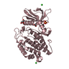
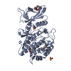
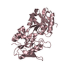
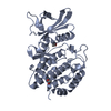
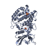
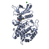
| Title | Structure of Human Vaccinia-related Kinase 1 (VRK1) bound to ACH471 | ||||||||||||
 Components Components | Serine/threonine-protein kinase VRK1 | ||||||||||||
 Keywords Keywords | TRANSFERASE/TRANSFERASE INHIBITOR /  Protein kinase / inhibitor / TRANSFERASE / TRANSFERASE-TRANSFERASE INHIBITOR complex Protein kinase / inhibitor / TRANSFERASE / TRANSFERASE-TRANSFERASE INHIBITOR complex | ||||||||||||
| Function / homology |  Function and homology information Function and homology informationGolgi disassembly / histone H3T3 kinase activity / Nuclear Envelope Breakdown / positive regulation of protein localization to chromatin / mitotic nuclear membrane disassembly / Golgi stack / Initiation of Nuclear Envelope (NE) Reformation / histone H3S10 kinase activity / kinase activity / histone binding ...Golgi disassembly / histone H3T3 kinase activity / Nuclear Envelope Breakdown / positive regulation of protein localization to chromatin / mitotic nuclear membrane disassembly / Golgi stack / Initiation of Nuclear Envelope (NE) Reformation / histone H3S10 kinase activity / kinase activity / histone binding / protein autophosphorylation / non-specific serine/threonine protein kinase / protein kinase activity / cell division / protein phosphorylation / protein serine kinase activity / protein serine/threonine kinase activity / DNA damage response / nucleolus / protein kinase binding / signal transduction / nucleoplasm / ATP binding / nucleus / cytosolSimilarity search - Function | ||||||||||||
| Biological species |  Homo sapiens (human) Homo sapiens (human) | ||||||||||||
| Method | X-RAY DIFFRACTION / SYNCHROTRON / MOLECULAR REPLACEMENT / Resolution: 2 Å | ||||||||||||
 Authors Authors | dos Reis, C.V. / Dutra, L.A. / Gama, F.H. / Mascarello, A. / Azevedo, H. / Guimaraes, C.R. / Massirer, K.B. / Arruda, P. / Edwards, A.M. / Counago, R.M. / Structural Genomics Consortium (SGC) | ||||||||||||
| Funding support |  Brazil, 3items Brazil, 3items
| ||||||||||||
 Citation Citation | Journal: To Be Published Title: Structure of Human Vaccinia-related Kinase 1 (VRK1) bound to ACH471 Authors: Guimaraes, C.R. / Counago, R.M. | ||||||||||||
| History |
|
- Structure visualization
| Structure viewer | Molecule: MolmilJmol/JSmol |
|---|
- Downloads & links
-Download
| PDBx/mmCIF format | 6vxu.cif.gz | 510 KB | Display | PDBx/mmCIF format |
|---|---|---|---|---|
| PDB format | pdb6vxu.ent.gz | 418.2 KB | Display | PDB format |
| PDBx/mmJSON format | 6vxu.json.gz | Tree view | PDBx/mmJSON format | |
| Others |  Other downloads Other downloads |
-Validation report
| Arichive directory | https://data.pdbj.org/pub/pdb/validation_reports/vx/6vxuftp://data.pdbj.org/pub/pdb/validation_reports/vx/6vxu | HTTPS FTP |
|---|
-Related structure data
| Related structure data |  6bruS S: Starting model for refinement |
|---|---|
| Similar structure data |


-Links
 PDBj
PDBj

- Assembly
| Deposited unit | 
| ||||||||
|---|---|---|---|---|---|---|---|---|---|
| 1 | 
| ||||||||
| 2 | 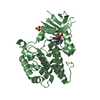
| ||||||||
| 3 | 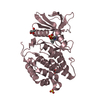
| ||||||||
| 4 | 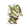
| ||||||||
| Unit cell |
|
-Components
-Protein , 1 types, 4 molecules ABCD
| #1: Protein | Mass: 41138.125 Da / Num. of mol.: 4 Mutation: K34A,K35A,E36A,E212A,K214A,E215A,E292A,K293A,K295A,K359A,K360A Source method: isolated from a genetically manipulated source Source: (gene. exp.) Homo sapiens (human) / Gene: VRK1 / Production host:  Escherichia coli BL21(DE3) (bacteria) / Strain (production host): BL21(DE3) Escherichia coli BL21(DE3) (bacteria) / Strain (production host): BL21(DE3)References: UniProt: Q99986, non-specific serine/threonine protein kinase |
|---|
-Non-polymers , 5 types, 609 molecules 


| #2: Chemical |  Mass: 399.394 Da / Num. of mol.: 3 / Source method: obtained synthetically / Formula: C20H19F2N5O2 / Feature type: SUBJECT OF INVESTIGATION Mass: 399.394 Da / Num. of mol.: 3 / Source method: obtained synthetically / Formula: C20H19F2N5O2 / Feature type: SUBJECT OF INVESTIGATION#3: Chemical | ChemComp-SO4 / Sulfate Mass: 96.063 Da / Num. of mol.: 14 / Source method: obtained synthetically / Formula: SO4 Mass: 96.063 Da / Num. of mol.: 14 / Source method: obtained synthetically / Formula: SO4#4: Chemical | ChemComp-VBD / ( |  Mass: 399.394 Da / Num. of mol.: 1 / Source method: obtained synthetically / Formula: C20H19F2N5O2 / Feature type: SUBJECT OF INVESTIGATION Mass: 399.394 Da / Num. of mol.: 1 / Source method: obtained synthetically / Formula: C20H19F2N5O2 / Feature type: SUBJECT OF INVESTIGATION#5: Chemical | ChemComp-GOL / | Glycerol Mass: 92.094 Da / Num. of mol.: 1 / Source method: obtained synthetically / Formula: C3H8O3 Mass: 92.094 Da / Num. of mol.: 1 / Source method: obtained synthetically / Formula: C3H8O3#6: Water | ChemComp-HOH / | WaterMass: 18.015 Da / Num. of mol.: 590 / Source method: isolated from a natural source / Formula: H2O |
|---|
-Details
| Has ligand of interest | Y |
|---|
-Experimental details
-Experiment
| Experiment | Method: X-RAY DIFFRACTION / Number of used crystals: 1 |
|---|
- Sample preparation
| Crystal | Density Matthews: 2.58 Å3/Da / Density % sol: 52.28 % |
|---|---|
| Crystal grow | Temperature: 293 K / Method: vapor diffusion, sitting drop / pH: 6.5 Details: 27.5% PEG3350; 300 mM LiSO4; 0.1 M SBG (each Sodium-tartrate + Bis-Tris + Glycylglycine) pH 6.5 |
-Data collection
| Diffraction | Mean temperature: 100 K / Serial crystal experiment: N |
|---|---|
| Diffraction source | Source: SYNCHROTRON / Site: APS  / Beamline: 24-ID-C / Wavelength: 0.97918 Å / Beamline: 24-ID-C / Wavelength: 0.97918 Å |
| Detector | Type: ADSC QUANTUM 315 / Detector: CCD / Date: Dec 5, 2019 |
| Radiation | Monochromator: Cryo-Cooled double crystal / Protocol: SINGLE WAVELENGTH / Monochromatic (M) / Laue (L): M / Scattering type: x-ray |
| Radiation wavelength | Wavelength: 0.97918 Å / Relative weight: 1 |
| Reflection | Resolution: 2→48.15 Å / Num. obs: 112736 / % possible obs: 97.8 % / Redundancy: 6.5 % / Rmerge(I) obs: 0.084 / Net I/σ(I): 11.9 |
| Reflection shell | Resolution: 2→2.03 Å / Redundancy: 4.5 % / Rmerge(I) obs: 0.944 / Num. unique obs: 4515 / % possible all: 80.8 |
- Processing
| Software |
| |||||||||||||||||||||||||||||||||||||||||||||||||||||||||||||||||||||||||||||||||||||||||||||||||||||||||||||||||||||||||||||||||||||||||||||||||
|---|---|---|---|---|---|---|---|---|---|---|---|---|---|---|---|---|---|---|---|---|---|---|---|---|---|---|---|---|---|---|---|---|---|---|---|---|---|---|---|---|---|---|---|---|---|---|---|---|---|---|---|---|---|---|---|---|---|---|---|---|---|---|---|---|---|---|---|---|---|---|---|---|---|---|---|---|---|---|---|---|---|---|---|---|---|---|---|---|---|---|---|---|---|---|---|---|---|---|---|---|---|---|---|---|---|---|---|---|---|---|---|---|---|---|---|---|---|---|---|---|---|---|---|---|---|---|---|---|---|---|---|---|---|---|---|---|---|---|---|---|---|---|---|---|---|---|
| Refinement | Method to determine structure: MOLECULAR REPLACEMENT Starting model: 6BRU Resolution: 2→30 Å / Cor.coef. Fo:Fc: 0.962 / Cor.coef. Fo:Fc free: 0.955 / SU B: 7.419 / SU ML: 0.101 / Cross valid method: THROUGHOUT / σ(F): 0 / ESU R: 0.152 / ESU R Free: 0.131 / Stereochemistry target values: MAXIMUM LIKELIHOOD Details: HYDROGENS HAVE BEEN ADDED IN THE RIDING POSITIONS U VALUES : WITH TLS ADDED
| |||||||||||||||||||||||||||||||||||||||||||||||||||||||||||||||||||||||||||||||||||||||||||||||||||||||||||||||||||||||||||||||||||||||||||||||||
| Solvent computation | Ion probe radii: 0.8 Å / Shrinkage radii: 0.8 Å / VDW probe radii: 1.2 Å / Solvent model: MASK | |||||||||||||||||||||||||||||||||||||||||||||||||||||||||||||||||||||||||||||||||||||||||||||||||||||||||||||||||||||||||||||||||||||||||||||||||
| Displacement parameters | Biso max: 118.71 Å2 / Biso mean: 39.26 Å2 / Biso min: 21.56 Å2
| |||||||||||||||||||||||||||||||||||||||||||||||||||||||||||||||||||||||||||||||||||||||||||||||||||||||||||||||||||||||||||||||||||||||||||||||||
| Refinement step | Cycle: final / Resolution: 2→30 Å
| |||||||||||||||||||||||||||||||||||||||||||||||||||||||||||||||||||||||||||||||||||||||||||||||||||||||||||||||||||||||||||||||||||||||||||||||||
| Refine LS restraints |
| |||||||||||||||||||||||||||||||||||||||||||||||||||||||||||||||||||||||||||||||||||||||||||||||||||||||||||||||||||||||||||||||||||||||||||||||||
| LS refinement shell | Resolution: 2→2.05 Å / Rfactor Rfree error: 0 / Total num. of bins used: 20
| |||||||||||||||||||||||||||||||||||||||||||||||||||||||||||||||||||||||||||||||||||||||||||||||||||||||||||||||||||||||||||||||||||||||||||||||||
| Refinement TLS params. | Method: refined / Refine-ID: X-RAY DIFFRACTION
| |||||||||||||||||||||||||||||||||||||||||||||||||||||||||||||||||||||||||||||||||||||||||||||||||||||||||||||||||||||||||||||||||||||||||||||||||
| Refinement TLS group |
|
