 Movie
Movie Controller

[English] 日本語
 Yorodumi
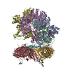
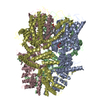
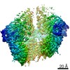
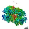
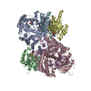
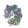
- PDB-6vvo: Structure of the human clamp loader (Replication Factor C, RFC) b... -
+ Open data

- Basic information
| Entry | Database: PDB / ID: 6vvo | ||||||||||||
|---|---|---|---|---|---|---|---|---|---|---|---|---|---|
| Title | Structure of the human clamp loader (Replication Factor C, RFC) bound to the sliding clamp (Proliferating Cell Nuclear Antigen, PCNA) | ||||||||||||
 Components Components |
| ||||||||||||
 Keywords Keywords |  DNA BINDING PROTEIN / REPLICATION / sliding clamp / DNA replication / AAA+ / ATPase / clamp loader DNA BINDING PROTEIN / REPLICATION / sliding clamp / DNA replication / AAA+ / ATPase / clamp loader | ||||||||||||
| Function / homology |  Function and homology information Function and homology informationDNA clamp unloader activity / positive regulation of deoxyribonuclease activity / dinucleotide insertion or deletion binding / PCNA-p21 complex / Elg1 RFC-like complex / DNA replication factor C complex / Ctf18 RFC-like complex / response to organophosphorus / mitotic telomere maintenance via semi-conservative replication / DNA clamp loader activity ...DNA clamp unloader activity / positive regulation of deoxyribonuclease activity / dinucleotide insertion or deletion binding / PCNA-p21 complex / Elg1 RFC-like complex / DNA replication factor C complex / Ctf18 RFC-like complex / response to organophosphorus / mitotic telomere maintenance via semi-conservative replication / DNA clamp loader activity / purine-specific mismatch base pair DNA N-glycosylase activity / positive regulation of DNA-directed DNA polymerase activity / MutLalpha complex binding / nuclear lamina / Polymerase switching / Telomere C-strand (Lagging Strand) Synthesis / Processive synthesis on the lagging strand / PCNA complex / Processive synthesis on the C-strand of the telomere / Removal of the Flap Intermediate / Mismatch repair (MMR) directed by MSH2:MSH3 (MutSbeta) / Mismatch repair (MMR) directed by MSH2:MSH6 (MutSalpha) / Polymerase switching on the C-strand of the telomere / Transcription of E2F targets under negative control by DREAM complex / Removal of the Flap Intermediate from the C-strand / replisome / DNA strand elongation involved in DNA replication / response to L-glutamate / DNA duplex unwinding / HDR through Single Strand Annealing (SSA) / Impaired BRCA2 binding to RAD51 / histone acetyltransferase binding / leading strand elongation / DNA synthesis involved in DNA repair / DNA polymerase processivity factor activity / replication fork processing / G1/S-Specific Transcription / response to dexamethasone / nuclear replication fork / Presynaptic phase of homologous DNA pairing and strand exchange / SUMOylation of DNA replication proteins / estrous cycle / telomere maintenance via telomerase / enzyme activator activity / PCNA-Dependent Long Patch Base Excision Repair / mismatch repair / cyclin-dependent protein kinase holoenzyme complex / translesion synthesis / ATP-dependent activity, acting on DNA / response to cadmium ion / Activation of ATR in response to replication stress / DNA polymerase binding / base-excision repair, gap-filling / positive regulation of DNA repair / epithelial cell differentiation / Translesion synthesis by REV1 / Translesion synthesis by POLK / Translesion synthesis by POLI / Gap-filling DNA repair synthesis and ligation in GG-NER / TP53 Regulates Transcription of Genes Involved in G2 Cell Cycle Arrest / replication fork / positive regulation of DNA replication / male germ cell nucleus / liver regeneration / nuclear estrogen receptor binding / Recognition of DNA damage by PCNA-containing replication complex / Termination of translesion DNA synthesis / Translesion Synthesis by POLH / HDR through Homologous Recombination (HRR) / G2/M DNA damage checkpoint / Dual Incision in GG-NER / DNA-templated DNA replication / receptor tyrosine kinase binding / cellular response to hydrogen peroxide / Dual incision in TC-NER / Gap-filling DNA repair synthesis and ligation in TC-NER / cellular response to UV / cellular response to xenobiotic stimulus / response to estradiol / E3 ubiquitin ligases ubiquitinate target proteins / heart development / Processing of DNA double-strand break ends / double-stranded DNA binding / Regulation of TP53 Activity through Phosphorylation / sequence-specific DNA binding / DNA replication / damaged DNA binding / chromosome, telomeric region / nuclear body / protein domain specific binding / DNA repair / centrosome / chromatin binding / chromatin / protein-containing complex binding / positive regulation of DNA-templated transcription / negative regulation of transcription by RNA polymerase II / enzyme binding / ATP hydrolysis activity / DNA bindingSimilarity search - Function | ||||||||||||
| Biological species |  Homo sapiens (human) Homo sapiens (human) | ||||||||||||
| Method | ELECTRON MICROSCOPY / single particle reconstruction / cryo EM / Resolution: 3.4 Å | ||||||||||||
 Authors Authors | Gaubitz, C. / Liu, X. / Stone, N.P. / Kelch, B.A. | ||||||||||||
| Funding support |  United States, United States,  Switzerland, 3items Switzerland, 3items
| ||||||||||||
 Citation Citation | Journal: Proc Natl Acad Sci U S A / Year: 2020 Title: Structure of the human clamp loader reveals an autoinhibited conformation of a substrate-bound AAA+ switch. Authors: Christl Gaubitz / Xingchen Liu / Joseph Magrino / Nicholas P Stone / Jacob Landeck / Mark Hedglin / Brian A Kelch / Abstract: DNA replication requires the sliding clamp, a ring-shaped protein complex that encircles DNA, where it acts as an essential cofactor for DNA polymerases and other proteins. The sliding clamp needs to ...DNA replication requires the sliding clamp, a ring-shaped protein complex that encircles DNA, where it acts as an essential cofactor for DNA polymerases and other proteins. The sliding clamp needs to be opened and installed onto DNA by a clamp loader ATPase of the AAA+ family. The human clamp loader replication factor C (RFC) and sliding clamp proliferating cell nuclear antigen (PCNA) are both essential and play critical roles in several diseases. Despite decades of study, no structure of human RFC has been resolved. Here, we report the structure of human RFC bound to PCNA by cryogenic electron microscopy to an overall resolution of ∼3.4 Å. The active sites of RFC are fully bound to adenosine 5'-triphosphate (ATP) analogs, which is expected to induce opening of the sliding clamp. However, we observe the complex in a conformation before PCNA opening, with the clamp loader ATPase modules forming an overtwisted spiral that is incapable of binding DNA or hydrolyzing ATP. The autoinhibited conformation observed here has many similarities to a previous yeast RFC:PCNA crystal structure, suggesting that eukaryotic clamp loaders adopt a similar autoinhibited state early on in clamp loading. Our results point to a "limited change/induced fit" mechanism in which the clamp first opens, followed by DNA binding, inducing opening of the loader to release autoinhibition. The proposed change from an overtwisted to an active conformation reveals an additional regulatory mechanism for AAA+ ATPases. Finally, our structural analysis of disease mutations leads to a mechanistic explanation for the role of RFC in human health. | ||||||||||||
| History |
|
- Structure visualization
| Movie |
Movie viewer |
|---|---|
| Structure viewer | Molecule: MolmilJmol/JSmol |
- Downloads & links
-Download
| PDBx/mmCIF format | 6vvo.cif.gz | 499.8 KB | Display | PDBx/mmCIF format |
|---|---|---|---|---|
| PDB format | pdb6vvo.ent.gz | 393 KB | Display | PDB format |
| PDBx/mmJSON format | 6vvo.json.gz | Tree view | PDBx/mmJSON format | |
| Others |  Other downloads Other downloads |
-Validation report
| Arichive directory | https://data.pdbj.org/pub/pdb/validation_reports/vv/6vvoftp://data.pdbj.org/pub/pdb/validation_reports/vv/6vvo | HTTPS FTP |
|---|
-Related structure data
| Related structure data | 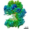 21405MC M: map data used to model this data C: citing same article ( |
|---|---|
| Similar structure data |

-Links
 PDBj
PDBj


- Assembly
| Deposited unit | 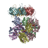
|
|---|---|
| 1 |
|
-Components
-Replication factor C subunit ... , 5 types, 5 molecules ABCDE
| #1: Protein | / Activator 1 140 kDa subunit / A1 140 kDa subunit / Activator 1 large subunit / Activator 1 subunit ...Activator 1 140 kDa subunit / A1 140 kDa subunit / Activator 1 large subunit / Activator 1 subunit 1 / DNA-binding protein PO-GA / Replication factor C 140 kDa subunit / RFC140 / Replication factor C large subunit Mass: 66221.227 Da / Num. of mol.: 1 / Fragment: UNP residues 556-1148 Source method: isolated from a genetically manipulated source Source: (gene. exp.) Homo sapiens (human) / Gene: RFC1, RFC140 / Production host:  Escherichia coli (E. coli) / References: UniProt: P35251 Escherichia coli (E. coli) / References: UniProt: P35251 |
|---|---|
| #2: Protein | / Activator 1 40 kDa subunit / A1 40 kDa subunit / Activator 1 subunit 2 / Replication factor C 40 ...Activator 1 40 kDa subunit / A1 40 kDa subunit / Activator 1 subunit 2 / Replication factor C 40 kDa subunit / RFC40 Mass: 39203.207 Da / Num. of mol.: 1 Source method: isolated from a genetically manipulated source Source: (gene. exp.) Homo sapiens (human) / Gene: RFC2 / Production host: Escherichia coli (E. coli) / References: UniProt: P35250 |
| #3: Protein | / Activator 1 36 kDa subunit / A1 36 kDa subunit / Activator 1 subunit 5 / Replication factor C 36 ...Activator 1 36 kDa subunit / A1 36 kDa subunit / Activator 1 subunit 5 / Replication factor C 36 kDa subunit / RFC36 Mass: 38545.512 Da / Num. of mol.: 1 Source method: isolated from a genetically manipulated source Source: (gene. exp.) Homo sapiens (human) / Gene: RFC5 / Production host: Escherichia coli (E. coli) / References: UniProt: P40937 |
| #4: Protein | / Activator 1 37 kDa subunit / A1 37 kDa subunit / Activator 1 subunit 4 / Replication factor C 37 ...Activator 1 37 kDa subunit / A1 37 kDa subunit / Activator 1 subunit 4 / Replication factor C 37 kDa subunit / RFC37 Mass: 39751.668 Da / Num. of mol.: 1 Source method: isolated from a genetically manipulated source Source: (gene. exp.) Homo sapiens (human) / Gene: RFC4 / Production host: Escherichia coli (E. coli) / References: UniProt: P35249 |
| #5: Protein | / Activator 1 38 kDa subunit / A1 38 kDa subunit / Activator 1 subunit 3 / Replication factor C 38 ...Activator 1 38 kDa subunit / A1 38 kDa subunit / Activator 1 subunit 3 / Replication factor C 38 kDa subunit / RFC38 Mass: 40614.332 Da / Num. of mol.: 1 Source method: isolated from a genetically manipulated source Source: (gene. exp.) Homo sapiens (human) / Gene: RFC3 / Production host: Escherichia coli (E. coli) / References: UniProt: P40938 |
-Protein , 1 types, 3 molecules FGH
| #6: Protein | / PCNA / Cyclin Mass: 28795.752 Da / Num. of mol.: 3 Source method: isolated from a genetically manipulated source Source: (gene. exp.) Homo sapiens (human) / Gene: PCNA / Production host: Escherichia coli (E. coli) / References: UniProt: P12004 |
|---|
-Non-polymers , 3 types, 9 molecules 


| #7: Chemical | ChemComp-MG /  Mass: 24.305 Da / Num. of mol.: 4 / Source method: obtained synthetically / Formula: Mg / Feature type: SUBJECT OF INVESTIGATION Mass: 24.305 Da / Num. of mol.: 4 / Source method: obtained synthetically / Formula: Mg / Feature type: SUBJECT OF INVESTIGATION#8: Chemical | ChemComp-AGS /  Mass: 523.247 Da / Num. of mol.: 4 / Source method: obtained synthetically / Formula: C10H16N5O12P3S / Feature type: SUBJECT OF INVESTIGATION / Comment: ATP-gamma-S, energy-carrying molecule analogue*YM Mass: 523.247 Da / Num. of mol.: 4 / Source method: obtained synthetically / Formula: C10H16N5O12P3S / Feature type: SUBJECT OF INVESTIGATION / Comment: ATP-gamma-S, energy-carrying molecule analogue*YM#9: Chemical | ChemComp-ADP / | Adenosine diphosphate Mass: 427.201 Da / Num. of mol.: 1 / Source method: obtained synthetically / Formula: C10H15N5O10P2 / Feature type: SUBJECT OF INVESTIGATION / Comment: ADP, energy-carrying molecule*YM Mass: 427.201 Da / Num. of mol.: 1 / Source method: obtained synthetically / Formula: C10H15N5O10P2 / Feature type: SUBJECT OF INVESTIGATION / Comment: ADP, energy-carrying molecule*YM |
|---|
-Details
| Has ligand of interest | Y |
|---|
-Experimental details
-Experiment
| Experiment | Method: ELECTRON MICROSCOPY |
|---|---|
| EM experiment | Aggregation state: PARTICLE / 3D reconstruction method: single particle reconstruction |
- Sample preparation
| Component | Name: Human Replication factor C (RFC) complex bound to the sliding clamp Proliferating cell nuclear antigen (PCNA) Type: COMPLEX Details: hRFC construct with a truncation of the A subunit's N-terminal region (missing residues 1-555) Entity ID: #1-#6 / Source: RECOMBINANT |
|---|---|
| Molecular weight | Value: 0.31 MDa / Experimental value: NO |
| Source (natural) | Organism: Homo sapiens (human) |
| Source (recombinant) | Organism: Escherichia coli (E. coli) |
| Buffer solution | pH: 7.5 |
| Specimen | Embedding applied: NO / Shadowing applied: NO / Staining applied: NO / Vitrification applied: YES |
| Specimen support | Grid material: GOLD / Grid type: Quantifoil R0.6/1 |
| Vitrification | Cryogen name: ETHANE / Humidity: 95 % / Chamber temperature: 283.15 K |
- Electron microscopy imaging
| Experimental equipment |  Model: Titan Krios / Image courtesy: FEI Company | |||||||||||||||||||||
|---|---|---|---|---|---|---|---|---|---|---|---|---|---|---|---|---|---|---|---|---|---|---|
| EM imaging | Accelerating voltage: 300 kV / Electron source
| |||||||||||||||||||||
| Image recording |
|
- Processing
| Software |
| ||||||||||||||||||||||||||||||||||||
|---|---|---|---|---|---|---|---|---|---|---|---|---|---|---|---|---|---|---|---|---|---|---|---|---|---|---|---|---|---|---|---|---|---|---|---|---|---|
| EM software |
| ||||||||||||||||||||||||||||||||||||
| CTF correction | Type: PHASE FLIPPING AND AMPLITUDE CORRECTION | ||||||||||||||||||||||||||||||||||||
| Symmetry | Point symmetry: C1 (asymmetric) | ||||||||||||||||||||||||||||||||||||
| 3D reconstruction | Resolution: 3.4 Å / Resolution method: FSC 0.143 CUT-OFF / Num. of particles: 193934 Details: Multi-body refinement was used to generate 2 reconstructions that were combined Symmetry type: POINT | ||||||||||||||||||||||||||||||||||||
| Atomic model building | Protocol: OTHER / Space: REAL Details: Initial local fitting was peformed using UCSF Chimera, followed by manual model adjustment, then refinement in PHENIX. | ||||||||||||||||||||||||||||||||||||
| Refinement | Cross valid method: NONE Stereochemistry target values: GeoStd + Monomer Library + CDL v1.2 | ||||||||||||||||||||||||||||||||||||
| Displacement parameters | Biso mean: 63.28 Å2 | ||||||||||||||||||||||||||||||||||||
| Refine LS restraints |
|
