 Movie
Movie Controller


+ Open data

- Basic information
| Entry | Database: PDB / ID: 6vpd | |||||||||||||||
|---|---|---|---|---|---|---|---|---|---|---|---|---|---|---|---|---|
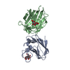
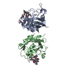
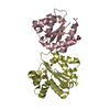
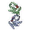
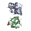
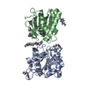
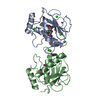
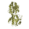
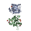
| Title | Crystal structure of Trgpx in apo form | |||||||||||||||
 Components Components | Glutathione peroxidase | |||||||||||||||
 Keywords Keywords | OXIDOREDUCTASE / glutathione peroxidase | |||||||||||||||
| Function / homology |  Function and homology information Function and homology information | |||||||||||||||
| Biological species |  Hypocrea jecorina (fungus) Hypocrea jecorina (fungus) | |||||||||||||||
| Method | X-RAY DIFFRACTION / SYNCHROTRON / MOLECULAR REPLACEMENT / Resolution: 2.603 Å | |||||||||||||||
 Authors Authors | Adriani, P.P. / De Oliveira, G.S. / Paiva, F.C.R. / Dias, M.V.B. / Chambergo, F.S. | |||||||||||||||
| Funding support |  Brazil, 4items Brazil, 4items
| |||||||||||||||
 Citation Citation | Journal: Int.J.Biol.Macromol. / Year: 2020 Title: Structural and functional characterization of the glutathione peroxidase-like thioredoxin peroxidase from the fungus Trichoderma reesei. Authors: Adriani, P.P. / de Paiva, F.C.R. / de Oliveira, G.S. / Leite, A.C. / Sanches, A.S. / Lopes, A.R. / Dias, M.V.B. / Chambergo, F.S. | |||||||||||||||
| History |
|
- Structure visualization
| Structure viewer | Molecule: MolmilJmol/JSmol |
|---|
- Downloads & links
-Download
| PDBx/mmCIF format | 6vpd.cif.gz | 73.8 KB | Display | PDBx/mmCIF format |
|---|---|---|---|---|
| PDB format | pdb6vpd.ent.gz | 53.1 KB | Display | PDB format |
| PDBx/mmJSON format | 6vpd.json.gz | Tree view | PDBx/mmJSON format | |
| Others |  Other downloads Other downloads |
-Validation report
| Arichive directory | https://data.pdbj.org/pub/pdb/validation_reports/vp/6vpdftp://data.pdbj.org/pub/pdb/validation_reports/vp/6vpd | HTTPS FTP |
|---|
-Related structure data
| Related structure data | 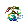 3cmiS S: Starting model for refinement |
|---|---|
| Similar structure data |

-Links
 PDBj
PDBj
- Assembly
| Deposited unit | 
| ||||||||
|---|---|---|---|---|---|---|---|---|---|
| 1 |
| ||||||||
| Unit cell |
|
-Components
| #1: Protein | Mass: 22197.305 Da / Num. of mol.: 2 Source method: isolated from a genetically manipulated source Source: (gene. exp.) Hypocrea jecorina (strain QM6a) (fungus)Strain: QM6a / Gene: TRIREDRAFT_47136 / Production host:  Escherichia coli BL21(DE3) (bacteria) / References: UniProt: G0RHF8 Escherichia coli BL21(DE3) (bacteria) / References: UniProt: G0RHF8#2: Water | ChemComp-HOH / | Water Mass: 18.015 Da / Num. of mol.: 14 / Source method: isolated from a natural source / Formula: H2O Mass: 18.015 Da / Num. of mol.: 14 / Source method: isolated from a natural source / Formula: H2O |
|---|
-Experimental details
-Experiment
| Experiment | Method: X-RAY DIFFRACTION / Number of used crystals: 1 |
|---|
- Sample preparation
| Crystal | Density Matthews: 1.98 Å3/Da / Density % sol: 37.93 % |
|---|---|
| Crystal grow | Temperature: 291.15 K / Method: vapor diffusion, hanging drop Details: 30% PEG 400, 200 mM magnesium chloride and 100 mM HEPES, pH7.5 |
-Data collection
| Diffraction | Mean temperature: 100 K / Serial crystal experiment: N |
|---|---|
| Diffraction source | Source: SYNCHROTRON / Site: PETRA III, EMBL c/o DESY  / Beamline: P13 (MX1) / Wavelength: 0.9763 Å / Beamline: P13 (MX1) / Wavelength: 0.9763 Å |
| Detector | Type: DECTRIS PILATUS 6M / Detector: PIXEL / Date: Nov 6, 2018 |
| Radiation | Protocol: SINGLE WAVELENGTH / Monochromatic (M) / Laue (L): M / Scattering type: x-ray |
| Radiation wavelength | Wavelength: 0.9763 Å / Relative weight: 1 |
| Reflection | Resolution: 2.6→39.88 Å / Num. obs: 10842 / % possible obs: 99.4 % / Redundancy: 4.7 % / Biso Wilson estimate: 43.52 Å2 / CC1/2: 0.977 / Rmerge(I) obs: 0.178 / Rpim(I) all: 0.087 / Rrim(I) all: 0.199 / Net I/σ(I): 7 / Num. measured all: 50721 / Scaling rejects: 100 |
| Reflection shell | Resolution: 2.6→2.72 Å / Redundancy: 4.7 % / Rmerge(I) obs: 0.746 / Num. measured all: 6150 / Num. unique obs: 1306 / CC1/2: 0.694 / Rpim(I) all: 0.372 / Rrim(I) all: 0.837 / Net I/σ(I) obs: 2.9 / % possible all: 98.6 |
- Processing
| Software |
| ||||||||||||||||||||||||||||||
|---|---|---|---|---|---|---|---|---|---|---|---|---|---|---|---|---|---|---|---|---|---|---|---|---|---|---|---|---|---|---|---|
| Refinement | Method to determine structure: MOLECULAR REPLACEMENT Starting model: 3cmi Resolution: 2.603→39.876 Å / SU ML: 0.27 / Cross valid method: THROUGHOUT / σ(F): 1.38 / Phase error: 25.28
| ||||||||||||||||||||||||||||||
| Solvent computation | Shrinkage radii: 0.9 Å / VDW probe radii: 1.11 Å | ||||||||||||||||||||||||||||||
| Displacement parameters | Biso max: 108.33 Å2 / Biso mean: 47.2477 Å2 / Biso min: 25.14 Å2 | ||||||||||||||||||||||||||||||
| Refinement step | Cycle: final / Resolution: 2.603→39.876 Å
| ||||||||||||||||||||||||||||||
| Refine LS restraints |
| ||||||||||||||||||||||||||||||
| LS refinement shell | Refine-ID: X-RAY DIFFRACTION / Rfactor Rfree error: 0
|
