 Movie
Movie Controller

[English] 日本語
 Yorodumi
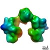
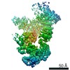
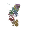
- PDB-6vp8: Cryo-EM structure of the C-terminal half of the Parkinson's Disea... -
+ Open data

- Basic information
| Entry | Database: PDB / ID: 6vp8 | ||||||||||||
|---|---|---|---|---|---|---|---|---|---|---|---|---|---|
| Title | Cryo-EM structure of the C-terminal half of the Parkinson's Disease-linked protein Leucine Rich Repeat Kinase 2 (LRRK2) | ||||||||||||
 Components Components | (Leucine-rich repeat serine/threonine-protein kinase 2) x 3 | ||||||||||||
 Keywords Keywords |  SIGNALING PROTEIN / Kinase / GTPase SIGNALING PROTEIN / Kinase / GTPase | ||||||||||||
| Function / homology |  Function and homology information Function and homology informationperoxidase inhibitor activity / caveola neck / negative regulation of thioredoxin peroxidase activity by peptidyl-threonine phosphorylation / negative regulation of protein processing involved in protein targeting to mitochondrion / Wnt signalosome assembly / beta-catenin destruction complex binding / regulation of branching morphogenesis of a nerve / regulation of kidney size / regulation of neuron maturation / tangential migration from the subventricular zone to the olfactory bulb ...peroxidase inhibitor activity / caveola neck / negative regulation of thioredoxin peroxidase activity by peptidyl-threonine phosphorylation / negative regulation of protein processing involved in protein targeting to mitochondrion / Wnt signalosome assembly / beta-catenin destruction complex binding / regulation of branching morphogenesis of a nerve / regulation of kidney size / regulation of neuron maturation / tangential migration from the subventricular zone to the olfactory bulb / protein localization to endoplasmic reticulum exit site / GTP-dependent protein kinase activity / regulation of neuroblast proliferation / regulation of ER to Golgi vesicle-mediated transport / regulation of synaptic vesicle transport / negative regulation of late endosome to lysosome transport / regulation of mitochondrial depolarization / negative regulation of protein targeting to mitochondrion / positive regulation of dopamine receptor signaling pathway / regulation of lysosomal lumen pH / regulation of CAMKK-AMPK signaling cascade / amphisome / mitochondrion localization / cytoplasmic side of mitochondrial outer membrane / multivesicular body, internal vesicle / co-receptor binding / regulation of retrograde transport, endosome to Golgi / negative regulation of excitatory postsynaptic potential / negative regulation of autophagosome assembly / regulation of dopamine receptor signaling pathway / positive regulation of microglial cell activation / neuron projection arborization / positive regulation of synaptic vesicle endocytosis / JUN kinase kinase kinase activity / olfactory bulb development / regulation of dendritic spine morphogenesis / regulation of protein kinase A signaling / striatum development / protein localization to mitochondrion / cellular response to dopamine / presynaptic cytosol / positive regulation of protein autoubiquitination / endoplasmic reticulum organization / Wnt signalosome / GTP metabolic process / positive regulation of programmed cell death / regulation of canonical Wnt signaling pathway / negative regulation of protein processing / syntaxin-1 binding / regulation of reactive oxygen species metabolic process / exploration behavior / negative regulation of GTPase activity / protein kinase A binding / regulation of locomotion / autolysosome / regulation of synaptic vesicle exocytosis / Golgi-associated vesicle / PTK6 promotes HIF1A stabilization / clathrin binding / negative regulation of macroautophagy / lysosome organization / regulation of mitochondrial fission / neuromuscular junction development / locomotory exploration behavior / intracellular distribution of mitochondria / Golgi organization / positive regulation of nitric-oxide synthase biosynthetic process / microvillus / Rho protein signal transduction / cellular response to organic cyclic compound / MAP kinase kinase kinase activity / canonical Wnt signaling pathway / positive regulation of protein kinase activity / cellular response to manganese ion / endoplasmic reticulum exit site / positive regulation of autophagy / negative regulation of endoplasmic reticulum stress-induced intrinsic apoptotic signaling pathway / JNK cascade / regulation of synaptic transmission, glutamatergic / excitatory postsynaptic potential / cellular response to starvation / dendrite cytoplasm / regulation of membrane potential / mitochondrion organization / GTPase activator activity / tubulin binding / SNARE binding / neuron projection morphogenesis / negative regulation of protein phosphorylation / negative regulation of protein binding / positive regulation of protein ubiquitination / regulation of autophagy / calcium-mediated signaling / determination of adult lifespan / mitochondrial membrane / Hydrolases; Acting on acid anhydrides; Acting on GTP to facilitate cellular and subcellular movement / peptidyl-threonine phosphorylation / regulation of protein stability / positive regulation of MAP kinase activity / trans-Golgi networkSimilarity search - Function | ||||||||||||
| Biological species |  Homo sapiens (human) Homo sapiens (human) | ||||||||||||
| Method | ELECTRON MICROSCOPY / single particle reconstruction / cryo EM / Resolution: 3.5 Å | ||||||||||||
 Authors Authors | Leschziner, A. / Deniston, C. / Lahiri, I. | ||||||||||||
| Funding support |  United States, 3items United States, 3items
| ||||||||||||
 Citation Citation | Journal: Nature / Year: 2020 Title: Structure of LRRK2 in Parkinson's disease and model for microtubule interaction. Authors: C K Deniston / J Salogiannis / S Mathea / D M Snead / I Lahiri / M Matyszewski / O Donosa / R Watanabe / J Böhning / A K Shiau / S Knapp / E Villa / S L Reck-Peterson / A E Leschziner /    Abstract: Leucine-rich repeat kinase 2 (LRRK2) is the most commonly mutated gene in familial Parkinson's disease and is also linked to its idiopathic form. LRRK2 has been proposed to function in membrane ...Leucine-rich repeat kinase 2 (LRRK2) is the most commonly mutated gene in familial Parkinson's disease and is also linked to its idiopathic form. LRRK2 has been proposed to function in membrane trafficking and colocalizes with microtubules. Despite the fundamental importance of LRRK2 for understanding and treating Parkinson's disease, structural information on the enzyme is limited. Here we report the structure of the catalytic half of LRRK2, and an atomic model of microtubule-associated LRRK2 built using a reported cryo-electron tomography in situ structure. We propose that the conformation of the LRRK2 kinase domain regulates its interactions with microtubules, with a closed conformation favouring oligomerization on microtubules. We show that the catalytic half of LRRK2 is sufficient for filament formation and blocks the motility of the microtubule-based motors kinesin 1 and cytoplasmic dynein 1 in vitro. Kinase inhibitors that stabilize an open conformation relieve this interference and reduce the formation of LRRK2 filaments in cells, whereas inhibitors that stabilize a closed conformation do not. Our findings suggest that LRRK2 can act as a roadblock for microtubule-based motors and have implications for the design of therapeutic LRRK2 kinase inhibitors. | ||||||||||||
| History |
|
- Structure visualization
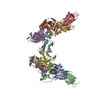
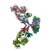
| Movie |
Movie viewer |
|---|---|
| Structure viewer | Molecule: MolmilJmol/JSmol |
- Downloads & links
-Download
| PDBx/mmCIF format | 6vp8.cif.gz | 333.5 KB | Display | PDBx/mmCIF format |
|---|---|---|---|---|
| PDB format | pdb6vp8.ent.gz | 266.5 KB | Display | PDB format |
| PDBx/mmJSON format | 6vp8.json.gz | Tree view | PDBx/mmJSON format | |
| Others |  Other downloads Other downloads |
-Validation report
| Arichive directory | https://data.pdbj.org/pub/pdb/validation_reports/vp/6vp8ftp://data.pdbj.org/pub/pdb/validation_reports/vp/6vp8 | HTTPS FTP |
|---|
-Related structure data
| Related structure data |  21250MC  6vnoC  6vp6C  6vp7C M: map data used to model this data C: citing same article ( |
|---|---|
| Similar structure data |


-Links
 PDBj
PDBj


- Assembly
| Deposited unit | 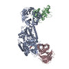
|
|---|---|
| 1 |
|
-Components
| #1: Protein | Mass: 136614.188 Da / Num. of mol.: 1 Source method: isolated from a genetically manipulated source Details: C-terminal residues 1330-2527 / Source: (gene. exp.) Homo sapiens (human) / Gene: LRRK2, PARK8 / Production host:   Spodoptera frugiperda (fall armyworm) Spodoptera frugiperda (fall armyworm)References: UniProt: Q5S007, non-specific serine/threonine protein kinase, Hydrolases; Acting on acid anhydrides; Acting on GTP to facilitate cellular and subcellular movement |
|---|---|
| #2: Protein | Mass: 32254.275 Da / Num. of mol.: 1 Source method: isolated from a genetically manipulated source Details: COR domain, residues 1670-1950 / Source: (gene. exp.) Homo sapiens (human) / Gene: LRRK2, PARK8 / Production host: Spodoptera frugiperda (fall armyworm)References: UniProt: Q5S007, non-specific serine/threonine protein kinase, Hydrolases; Acting on acid anhydrides; Acting on GTP to facilitate cellular and subcellular movement |
| #3: Protein | Mass: 40178.562 Da / Num. of mol.: 1 Source method: isolated from a genetically manipulated source Details: WD40 domain, residues 2140-2498 / Source: (gene. exp.) Homo sapiens (human) / Gene: LRRK2 / Production host: Spodoptera frugiperda (fall armyworm)References: UniProt: Q5S007, non-specific serine/threonine protein kinase, Hydrolases; Acting on acid anhydrides; Acting on GTP to facilitate cellular and subcellular movement |
| Has ligand of interest | Y |
-Experimental details
-Experiment
| Experiment | Method: ELECTRON MICROSCOPY |
|---|---|
| EM experiment | Aggregation state: PARTICLE / 3D reconstruction method: single particle reconstruction |
- Sample preparation
| Component | Name: The C-terminal half of the Leucine Rich Repeat Kinase 2 (LRRK2) protein Type: COMPLEX / Entity ID: all / Source: RECOMBINANT | |||||||||||||||||||||||||||||||||||
|---|---|---|---|---|---|---|---|---|---|---|---|---|---|---|---|---|---|---|---|---|---|---|---|---|---|---|---|---|---|---|---|---|---|---|---|---|
| Molecular weight | Value: 0.137 MDa / Experimental value: NO | |||||||||||||||||||||||||||||||||||
| Source (natural) | Organism: Homo sapiens (human) | |||||||||||||||||||||||||||||||||||
| Source (recombinant) | Organism: Spodoptera frugiperda (fall armyworm) | |||||||||||||||||||||||||||||||||||
| Buffer solution | pH: 7.4 | |||||||||||||||||||||||||||||||||||
| Buffer component |
| |||||||||||||||||||||||||||||||||||
| Specimen | Conc.: 0.5 mg/ml / Embedding applied: NO / Shadowing applied: NO / Staining applied: NO / Vitrification applied: YES / Details: 4uM concentration | |||||||||||||||||||||||||||||||||||
| Specimen support | Grid material: GOLD / Grid type: Quantifoil, UltrAuFoil, R1.2/1.3 | |||||||||||||||||||||||||||||||||||
| Vitrification | Instrument: FEI VITROBOT MARK II / Cryogen name: ETHANE / Humidity: 100 % / Chamber temperature: 277.15 K |
- Electron microscopy imaging
| Experimental equipment |  Model: Titan Krios / Image courtesy: FEI Company |
|---|---|
| Microscopy | Model: FEI TITAN KRIOS |
| Electron gun | Electron source: FIELD EMISSION GUN / Accelerating voltage: 300 kV / Illumination mode: FLOOD BEAM |
| Electron lens | Mode: BRIGHT FIELDBright-field microscopy / Nominal magnification: 130000 X / Nominal defocus max: 1800 nm / Nominal defocus min: 1000 nm / Cs: 2.7 mm |
| Specimen holder | Cryogen: NITROGEN / Specimen holder model: FEI TITAN KRIOS AUTOGRID HOLDER |
| Image recording | Average exposure time: 8 sec. / Electron dose: 6.65 e/Å2 / Detector mode: COUNTING / Film or detector model: GATAN K2 SUMMIT (4k x 4k) / Num. of grids imaged: 1 / Num. of real images: 3826 |
| EM imaging optics | Energyfilter name: GIF 2002 |
| Image scans | Movie frames/image: 40 |
- Processing
| EM software |
| |||||||||||||||||||||||||||||||||||||||||||||||||||||||||||||||||
|---|---|---|---|---|---|---|---|---|---|---|---|---|---|---|---|---|---|---|---|---|---|---|---|---|---|---|---|---|---|---|---|---|---|---|---|---|---|---|---|---|---|---|---|---|---|---|---|---|---|---|---|---|---|---|---|---|---|---|---|---|---|---|---|---|---|---|
| CTF correction | Details: Per-particle CTF values / Type: PHASE FLIPPING AND AMPLITUDE CORRECTION | |||||||||||||||||||||||||||||||||||||||||||||||||||||||||||||||||
| Particle selection | Num. of particles selected: 836956 | |||||||||||||||||||||||||||||||||||||||||||||||||||||||||||||||||
| Symmetry | Point symmetry: C3 (3 fold cyclic) | |||||||||||||||||||||||||||||||||||||||||||||||||||||||||||||||||
| 3D reconstruction | Resolution: 3.5 Å / Resolution method: FSC 0.143 CUT-OFF / Num. of particles: 70953 Details: For the signal subtracted map, 105,787 particles went into the final map that achieved 3.8A resolution Symmetry type: POINT |
