 Movie
Movie Controller

[English] 日本語
 Yorodumi
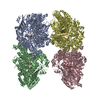
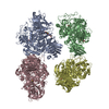
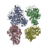
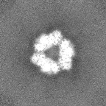
- EMDB-21250: Cryo-EM structure of the C-terminal half of the Parkinson's Disea... -
+ Open data

- Basic information
| Entry | Database: EMDB / ID: EMD-21250 | ||||||||||||
|---|---|---|---|---|---|---|---|---|---|---|---|---|---|
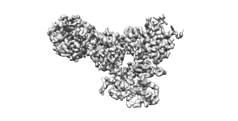
| Title | Cryo-EM structure of the C-terminal half of the Parkinson's Disease-linked protein Leucine Rich Repeat Kinase 2 (LRRK2) | ||||||||||||
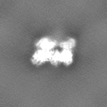
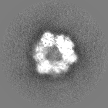
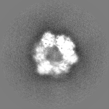
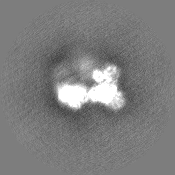
 Map data Map data | 3.8A signal subtracted locally filtered cryo-EM map of C-terminal half of Leucine Rich Repeat Kinase 2 (LRRK2) | ||||||||||||
 Sample Sample |
| ||||||||||||
| Function / homology |  Function and homology information Function and homology informationperoxidase inhibitor activity / caveola neck / negative regulation of thioredoxin peroxidase activity by peptidyl-threonine phosphorylation / negative regulation of protein processing involved in protein targeting to mitochondrion / Wnt signalosome assembly / beta-catenin destruction complex binding / regulation of branching morphogenesis of a nerve / regulation of kidney size / regulation of neuron maturation / tangential migration from the subventricular zone to the olfactory bulb ...peroxidase inhibitor activity / caveola neck / negative regulation of thioredoxin peroxidase activity by peptidyl-threonine phosphorylation / negative regulation of protein processing involved in protein targeting to mitochondrion / Wnt signalosome assembly / beta-catenin destruction complex binding / regulation of branching morphogenesis of a nerve / regulation of kidney size / regulation of neuron maturation / tangential migration from the subventricular zone to the olfactory bulb / protein localization to endoplasmic reticulum exit site / GTP-dependent protein kinase activity / regulation of neuroblast proliferation / regulation of ER to Golgi vesicle-mediated transport / regulation of synaptic vesicle transport / negative regulation of late endosome to lysosome transport / regulation of mitochondrial depolarization / negative regulation of protein targeting to mitochondrion / positive regulation of dopamine receptor signaling pathway / regulation of lysosomal lumen pH / regulation of CAMKK-AMPK signaling cascade / amphisome / mitochondrion localization / cytoplasmic side of mitochondrial outer membrane /  co-receptor binding / regulation of retrograde transport, endosome to Golgi / negative regulation of excitatory postsynaptic potential / negative regulation of autophagosome assembly / regulation of dopamine receptor signaling pathway / positive regulation of microglial cell activation / neuron projection arborization / positive regulation of synaptic vesicle endocytosis / JUN kinase kinase kinase activity / olfactory bulb development / regulation of dendritic spine morphogenesis / regulation of protein kinase A signaling / multivesicular body, internal vesicle / striatum development / protein localization to mitochondrion / cellular response to dopamine / presynaptic cytosol / positive regulation of protein autoubiquitination / endoplasmic reticulum organization / Wnt signalosome / positive regulation of programmed cell death / GTP metabolic process / regulation of canonical Wnt signaling pathway / negative regulation of protein processing / syntaxin-1 binding / regulation of reactive oxygen species metabolic process / negative regulation of GTPase activity / regulation of locomotion / autolysosome / protein kinase A binding / exploration behavior / regulation of synaptic vesicle exocytosis / Golgi-associated vesicle / PTK6 promotes HIF1A stabilization / clathrin binding / negative regulation of macroautophagy / lysosome organization / regulation of mitochondrial fission / neuromuscular junction development / intracellular distribution of mitochondria / Golgi organization / positive regulation of nitric-oxide synthase biosynthetic process / locomotory exploration behavior / microvillus / Rho protein signal transduction / cellular response to organic cyclic compound / MAP kinase kinase kinase activity / canonical Wnt signaling pathway / positive regulation of protein kinase activity / cellular response to manganese ion / endoplasmic reticulum exit site / negative regulation of endoplasmic reticulum stress-induced intrinsic apoptotic signaling pathway / positive regulation of autophagy / JNK cascade / regulation of synaptic transmission, glutamatergic / excitatory postsynaptic potential / regulation of membrane potential / cellular response to starvation / dendrite cytoplasm / GTPase activator activity / mitochondrion organization / tubulin binding / SNARE binding / neuron projection morphogenesis / negative regulation of protein phosphorylation / negative regulation of protein binding / positive regulation of protein ubiquitination / regulation of autophagy / determination of adult lifespan / calcium-mediated signaling / mitochondrial membrane / Hydrolases; Acting on acid anhydrides; Acting on GTP to facilitate cellular and subcellular movement / peptidyl-threonine phosphorylation / regulation of protein stability / positive regulation of MAP kinase activity / trans-Golgi network co-receptor binding / regulation of retrograde transport, endosome to Golgi / negative regulation of excitatory postsynaptic potential / negative regulation of autophagosome assembly / regulation of dopamine receptor signaling pathway / positive regulation of microglial cell activation / neuron projection arborization / positive regulation of synaptic vesicle endocytosis / JUN kinase kinase kinase activity / olfactory bulb development / regulation of dendritic spine morphogenesis / regulation of protein kinase A signaling / multivesicular body, internal vesicle / striatum development / protein localization to mitochondrion / cellular response to dopamine / presynaptic cytosol / positive regulation of protein autoubiquitination / endoplasmic reticulum organization / Wnt signalosome / positive regulation of programmed cell death / GTP metabolic process / regulation of canonical Wnt signaling pathway / negative regulation of protein processing / syntaxin-1 binding / regulation of reactive oxygen species metabolic process / negative regulation of GTPase activity / regulation of locomotion / autolysosome / protein kinase A binding / exploration behavior / regulation of synaptic vesicle exocytosis / Golgi-associated vesicle / PTK6 promotes HIF1A stabilization / clathrin binding / negative regulation of macroautophagy / lysosome organization / regulation of mitochondrial fission / neuromuscular junction development / intracellular distribution of mitochondria / Golgi organization / positive regulation of nitric-oxide synthase biosynthetic process / locomotory exploration behavior / microvillus / Rho protein signal transduction / cellular response to organic cyclic compound / MAP kinase kinase kinase activity / canonical Wnt signaling pathway / positive regulation of protein kinase activity / cellular response to manganese ion / endoplasmic reticulum exit site / negative regulation of endoplasmic reticulum stress-induced intrinsic apoptotic signaling pathway / positive regulation of autophagy / JNK cascade / regulation of synaptic transmission, glutamatergic / excitatory postsynaptic potential / regulation of membrane potential / cellular response to starvation / dendrite cytoplasm / GTPase activator activity / mitochondrion organization / tubulin binding / SNARE binding / neuron projection morphogenesis / negative regulation of protein phosphorylation / negative regulation of protein binding / positive regulation of protein ubiquitination / regulation of autophagy / determination of adult lifespan / calcium-mediated signaling / mitochondrial membrane / Hydrolases; Acting on acid anhydrides; Acting on GTP to facilitate cellular and subcellular movement / peptidyl-threonine phosphorylation / regulation of protein stability / positive regulation of MAP kinase activity / trans-Golgi networkSimilarity search - Function | ||||||||||||
| Biological species |  Homo sapiens (human) Homo sapiens (human) | ||||||||||||
| Method | single particle reconstruction / cryo EM / Resolution: 3.5 Å | ||||||||||||
 Authors Authors | Leschziner A / Deniston C / Lahiri I | ||||||||||||
| Funding support |  United States, 3 items United States, 3 items
| ||||||||||||
 Citation Citation | Journal: Nature / Year: 2020 Title: Structure of LRRK2 in Parkinson's disease and model for microtubule interaction. Authors: C K Deniston / J Salogiannis / S Mathea / D M Snead / I Lahiri / M Matyszewski / O Donosa / R Watanabe / J Böhning / A K Shiau / S Knapp / E Villa / S L Reck-Peterson / A E Leschziner /    Abstract: Leucine-rich repeat kinase 2 (LRRK2) is the most commonly mutated gene in familial Parkinson's disease and is also linked to its idiopathic form. LRRK2 has been proposed to function in membrane ...Leucine-rich repeat kinase 2 (LRRK2) is the most commonly mutated gene in familial Parkinson's disease and is also linked to its idiopathic form. LRRK2 has been proposed to function in membrane trafficking and colocalizes with microtubules. Despite the fundamental importance of LRRK2 for understanding and treating Parkinson's disease, structural information on the enzyme is limited. Here we report the structure of the catalytic half of LRRK2, and an atomic model of microtubule-associated LRRK2 built using a reported cryo-electron tomography in situ structure. We propose that the conformation of the LRRK2 kinase domain regulates its interactions with microtubules, with a closed conformation favouring oligomerization on microtubules. We show that the catalytic half of LRRK2 is sufficient for filament formation and blocks the motility of the microtubule-based motors kinesin 1 and cytoplasmic dynein 1 in vitro. Kinase inhibitors that stabilize an open conformation relieve this interference and reduce the formation of LRRK2 filaments in cells, whereas inhibitors that stabilize a closed conformation do not. Our findings suggest that LRRK2 can act as a roadblock for microtubule-based motors and have implications for the design of therapeutic LRRK2 kinase inhibitors. | ||||||||||||
| History |
|
- Structure visualization
| Movie |
Movie viewer |
|---|---|
| Structure viewer | EM map: SurfViewMolmilJmol/JSmol |
| Supplemental images |
- Downloads & links
-EMDB archive
| Map data | emd_21250.map.gz | 100.9 MB | EMDB map data format | |
|---|---|---|---|---|
| Header (meta data) | emd-21250-v30.xmlemd-21250.xml | 28.3 KB 28.3 KB | Display Display | EMDB header |
| FSC (resolution estimation) | emd_21250_fsc_1.xmlemd_21250_fsc_2.xml | 12.7 KB 12.7 KB | Display Display | FSC data file |
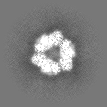
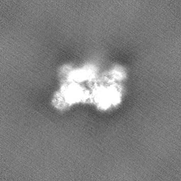
| Images |  emd_21250.png emd_21250.png | 154 KB | ||
| Others | emd_21250_additional_1.map.gzemd_21250_additional_2.map.gzemd_21250_additional_3.map.gzemd_21250_half_map_1.map.gzemd_21250_half_map_2.map.gz | 162.5 MB 159.6 MB 159.6 MB 136.1 MB 136 MB | ||
| Archive directory |  http://ftp.pdbj.org/pub/emdb/structures/EMD-21250ftp://ftp.pdbj.org/pub/emdb/structures/EMD-21250 http://ftp.pdbj.org/pub/emdb/structures/EMD-21250ftp://ftp.pdbj.org/pub/emdb/structures/EMD-21250 | HTTPS FTP |
-Related structure data
| Related structure data |  6vnoMC  6vp6MC  6vp7MC  6vp8MC M: atomic model generated by this map C: citing same article ( |
|---|---|
| Similar structure data |


-Links
| EMDB pages | EMDB (EBI/PDBe) / EMDataResource |
|---|---|
| Related items in Molecule of the Month |


-Map
| File | Download / File: emd_21250.map.gz / Format: CCP4 / Size: 172.1 MB / Type: IMAGE STORED AS FLOATING POINT NUMBER (4 BYTES) | ||||||||||||||||||||||||||||||||||||||||||||||||||||||||||||
|---|---|---|---|---|---|---|---|---|---|---|---|---|---|---|---|---|---|---|---|---|---|---|---|---|---|---|---|---|---|---|---|---|---|---|---|---|---|---|---|---|---|---|---|---|---|---|---|---|---|---|---|---|---|---|---|---|---|---|---|---|---|
| Annotation | 3.8A signal subtracted locally filtered cryo-EM map of C-terminal half of Leucine Rich Repeat Kinase 2 (LRRK2) | ||||||||||||||||||||||||||||||||||||||||||||||||||||||||||||
| Voxel size | X=Y=Z: 1.07 Å | ||||||||||||||||||||||||||||||||||||||||||||||||||||||||||||
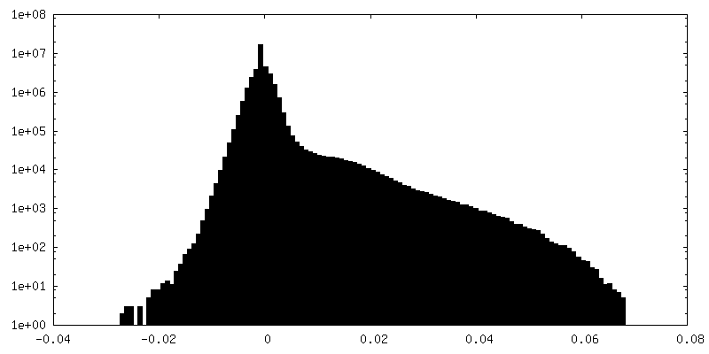
| Density |
| ||||||||||||||||||||||||||||||||||||||||||||||||||||||||||||
| Symmetry | Space group: 0 | ||||||||||||||||||||||||||||||||||||||||||||||||||||||||||||
| Details | EMDB XML:
CCP4 map header:
| ||||||||||||||||||||||||||||||||||||||||||||||||||||||||||||
-Supplemental data
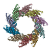
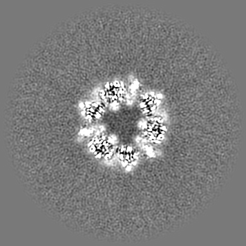
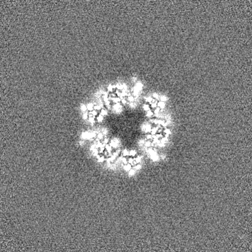
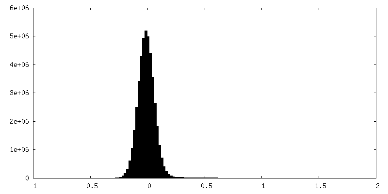
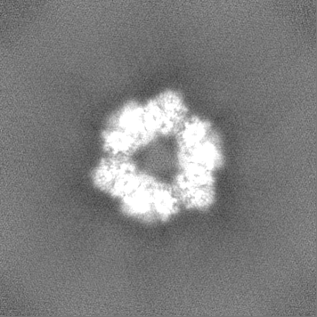
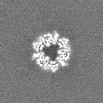
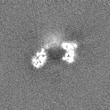
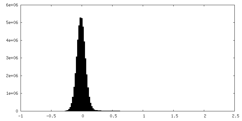
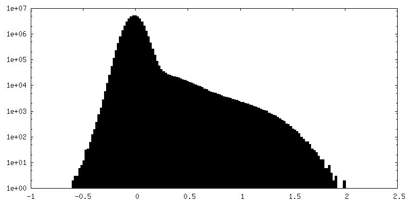
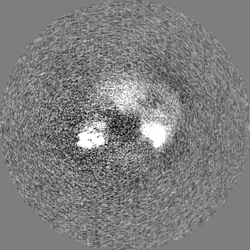
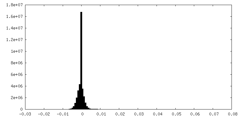
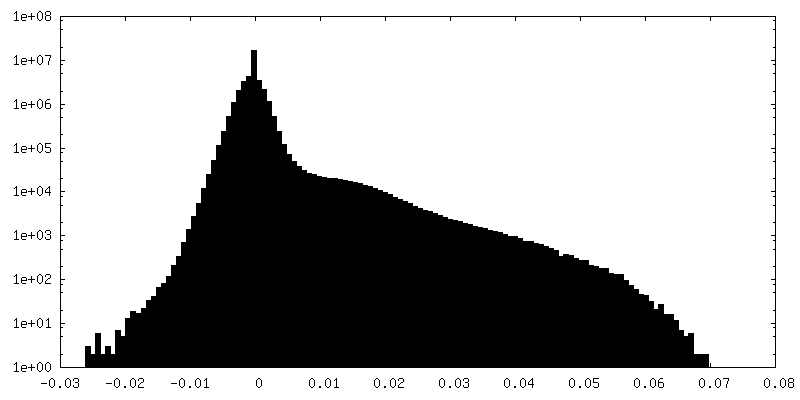
-Additional map: 3.47A C3 symmetric locally filtered cryo-EM map of...
| File | emd_21250_additional_1.map | ||||||||||||
|---|---|---|---|---|---|---|---|---|---|---|---|---|---|
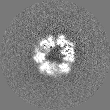
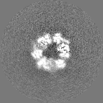
| Annotation | 3.47A C3 symmetric locally filtered cryo-EM map of C-terminal half of Leucine Rich Repeat Kinase 2 (LRRK2). | ||||||||||||
| Projections & Slices |
| ||||||||||||
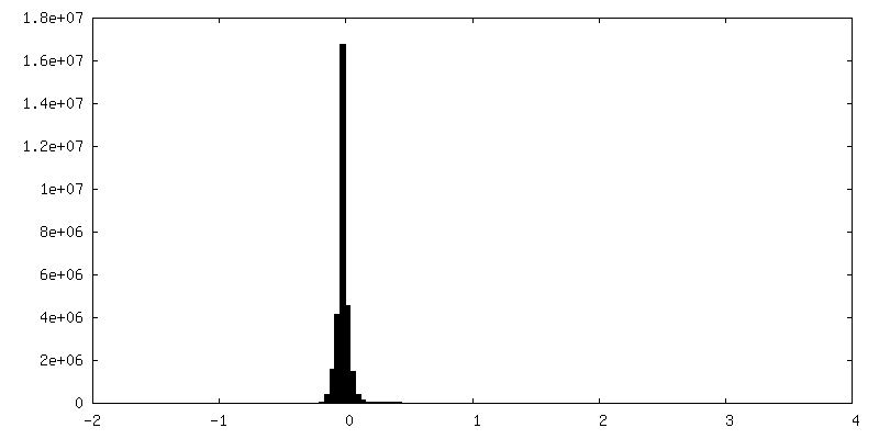
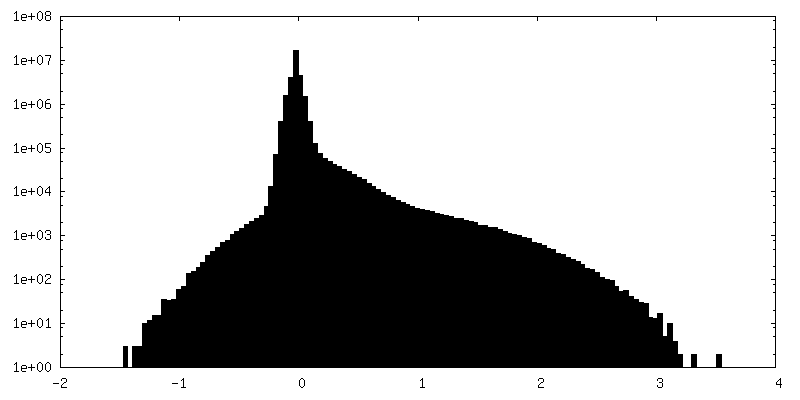
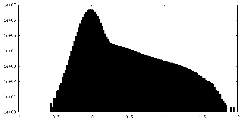
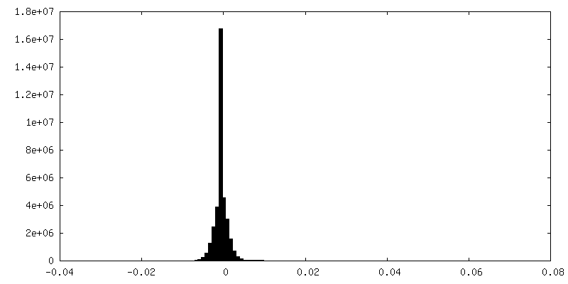
| Density Histograms |
 Z
Z Y
Y X
X


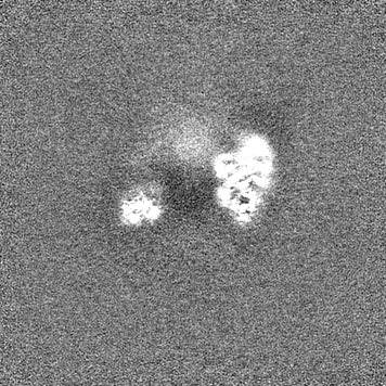
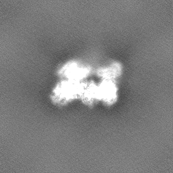
-Additional map: Half map 1 of C3 symmetric map
| File | emd_21250_additional_2.map | ||||||||||||
|---|---|---|---|---|---|---|---|---|---|---|---|---|---|
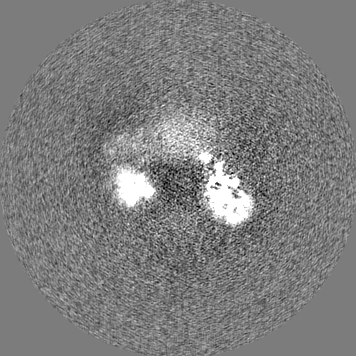
| Annotation | Half map 1 of C3 symmetric map | ||||||||||||
| Projections & Slices |
| ||||||||||||
| Density Histograms |

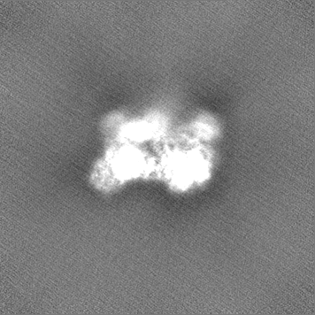
-Additional map: Half map 2 of C3 symmetric map
| File | emd_21250_additional_3.map | ||||||||||||
|---|---|---|---|---|---|---|---|---|---|---|---|---|---|
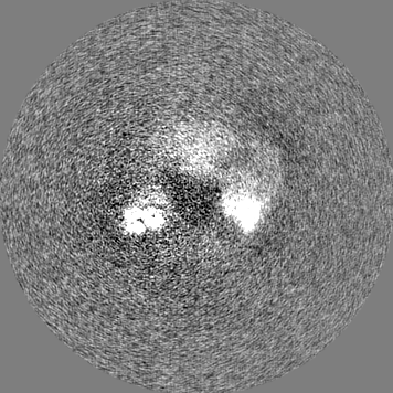
| Annotation | Half map 2 of C3 symmetric map | ||||||||||||
| Projections & Slices |
| ||||||||||||
| Density Histograms |

-Half map: Half map 1 of signal subtracted map
| File | emd_21250_half_map_1.map | ||||||||||||
|---|---|---|---|---|---|---|---|---|---|---|---|---|---|
| Annotation | Half map 1 of signal subtracted map | ||||||||||||
| Projections & Slices |
| ||||||||||||
| Density Histograms |

-Half map: Half map 2 of signal subtracted map
| File | emd_21250_half_map_2.map | ||||||||||||
|---|---|---|---|---|---|---|---|---|---|---|---|---|---|
| Annotation | Half map 2 of signal subtracted map | ||||||||||||
| Projections & Slices |
| ||||||||||||
| Density Histograms |

- Sample components
-Entire : The C-terminal half of the Leucine Rich Repeat Kinase 2 (LRRK2) p...
| Entire | Name: The C-terminal half of the Leucine Rich Repeat Kinase 2 (LRRK2) protein. |
|---|---|
| Components |
|
-Supramolecule #1: The C-terminal half of the Leucine Rich Repeat Kinase 2 (LRRK2) p...
| Supramolecule | Name: The C-terminal half of the Leucine Rich Repeat Kinase 2 (LRRK2) protein. type: complex / ID: 1 / Parent: 0 / Macromolecule list: #1 / Details: C-terminal half runs from residue 1327-2527. |
|---|---|
| Source (natural) | Organism: Homo sapiens (human) |
| Recombinant expression | Organism:   Spodoptera frugiperda (fall armyworm) Spodoptera frugiperda (fall armyworm) |
| Molecular weight | Theoretical: 137 KDa |
-Macromolecule #1: Leucine-rich repeat serine/threonine-protein kinase 2
| Macromolecule | Name: Leucine-rich repeat serine/threonine-protein kinase 2 / type: protein_or_peptide / ID: 1 / Details: C-terminal residues 1330-2527 / Number of copies: 1 / Enantiomer: LEVO / EC number: non-specific serine/threonine protein kinase |
|---|---|
| Source (natural) | Organism: Homo sapiens (human) |
| Molecular weight | Theoretical: 136.943609 KDa |
| Recombinant expression | Organism: Spodoptera frugiperda (fall armyworm) |
| Sequence | String: KKAVPYNRMK LMIVGN(TPO)GSG KTTLLQQLMK TKKSDLGMQS ATVGIDVKDW PIQIRDKRKR DLVLNVWDFA GREEFY STH PHFMTQRALY LAVYDLSKGQ AEVDAMKPWL FNIKARASSS PVILVGTHLD VSDEKQRKAC MSKITKELLN KRGFPAI RD YHFVNATEES ...String: KKAVPYNRMK LMIVGN(TPO)GSG KTTLLQQLMK TKKSDLGMQS ATVGIDVKDW PIQIRDKRKR DLVLNVWDFA GREEFY STH PHFMTQRALY LAVYDLSKGQ AEVDAMKPWL FNIKARASSS PVILVGTHLD VSDEKQRKAC MSKITKELLN KRGFPAI RD YHFVNATEES DALAKLRKTI INESLNFKIR DQLVVGQLIP DCYVELEKII LSERKNVPIE FPVIDRKRLL QLVRENQL Q LDENELPHAV HFLNESGVLL HFQDPALQLS DLYFVEPKWL CKIMAQILTV KVEGCPKHPK GIISRRDVEK FLSKKRKFP KNYMSQYFKL LEKFQIALPI GEEYLLVPSS LSDHRPVIEL PHCENSEIII RLYEMPYFPM GFWSRLINRL LEISPYMLSG RERALRPNR MYWRQGIYLN WSPEAYCLVG SEVLDNHPES FLKITVPSCR KGCILLGQVV DHIDSLMEEW FPGLLEIDIC G EGETLLKK WALYSFNDGE EHQKILLDDL MKKAEEGDLL VNPDQPRLTI PISQIAPDLI LADLPRNIML NNDELEFEQA PE FLLGDGS FGSVYRAAYE GEEVAVKIFN KHTSLRLLRQ ELVVLCHLHH PSLISLLAAG IRPRMLVMEL ASKGSLDRLL QQD KASLTR TLQHRIALHV ADGLRYLHSA MIIYRDLKPH NVLLFTLYPN AAIIAKIADY GIAQYCCRMG IKTSEGTPGF RAPE VARGN VIYNQQADVY SFGLLLYDIL TTGGRIVEGL KFPNEFDELE IQGKLPDPVK EYGCAPWPMV EKLIKQCLKE NPQER PTSA QVFDILNSAE LVCLTRRILL PKNVIVECMV ATHHNSRNAS IWLGCGHTDR GQLSFLDLNT EGYTSEEVAD SRILCL ALV HLPVEKESWI VSGTQSGTLL VINTEDGKKR HTLEKMTDSV TCLYCNSFSK QSKQKNFLLV GTADGKLAIF EDKTVKL KG AAPLKILNIG NVSTPLMCLS ESTNSTERNV MWGGCGTKIF SFSNDFTIQK LIETRTSQLF SYAAFSDSNI ITVVVDTA L YIAKQNSPVV EVWDKKTEKL CGLIDCVHFL REVMVKENKE SKHKMSYSGR VKTLCLQKNT ALWIGTGGGH ILLLDLSTR RLIRVIYNFC NSVRVMMTAQ LGSLKNVMLV LGYNRKNTEG TQKQKEIQSC LTVWDINLPH EVQNLEKHIE VRKELAEKMR RTSVE |
-Macromolecule #2: GUANOSINE-5'-DIPHOSPHATE
| Macromolecule | Name: GUANOSINE-5'-DIPHOSPHATE / type: ligand / ID: 2 / Number of copies: 1 / Formula: GDP |
|---|---|
| Molecular weight | Theoretical: 443.201 Da |
| Chemical component information |  ChemComp-GDP: |
-Macromolecule #3: MAGNESIUM ION
| Macromolecule | Name: MAGNESIUM ION / type: ligand / ID: 3 / Number of copies: 1 / Formula: MG |
|---|---|
| Molecular weight | Theoretical: 24.305 Da |
-Experimental details
-Structure determination
| Method | cryo EM |
|---|---|
 Processing Processing | single particle reconstruction |
| Aggregation state | particle |
-Sample preparation
| Concentration | 0.5 mg/mL | |||||||||||||||||||||
|---|---|---|---|---|---|---|---|---|---|---|---|---|---|---|---|---|---|---|---|---|---|---|
| Buffer | pH: 7.4 Component:
| |||||||||||||||||||||
| Grid | Model: Quantifoil, UltrAuFoil, R1.2/1.3 / Material: GOLD / Pretreatment - Type: GLOW DISCHARGE | |||||||||||||||||||||
| Vitrification | Cryogen name: ETHANE / Chamber humidity: 100 % / Chamber temperature: 277 K / Instrument: FEI VITROBOT MARK II | |||||||||||||||||||||
| Details | 4uM concentration |
- Electron microscopy
| Microscope | FEI TITAN KRIOS |
|---|---|
| Electron beam | Acceleration voltage: 300 kV / Electron source: FIELD EMISSION GUN |
| Electron optics | Illumination mode: FLOOD BEAM / Imaging mode: BRIGHT FIELDBright-field microscopy / Cs: 2.7 mm / Nominal defocus max: 1.8 µm / Nominal defocus min: 1.0 µm / Nominal magnification: 130000 |
| Specialist optics | Energy filter - Name: GIF 2002 |
| Sample stage | Specimen holder model: FEI TITAN KRIOS AUTOGRID HOLDER / Cooling holder cryogen: NITROGEN |
| Image recording | Film or detector model: GATAN K2 SUMMIT (4k x 4k) / Detector mode: COUNTING / Number grids imaged: 1 / Number real images: 3826 / Average exposure time: 8.0 sec. / Average electron dose: 6.65 e/Å2 |
| Experimental equipment |  Model: Titan Krios / Image courtesy: FEI Company |
-Image processing
| Particle selection | Number selected: 836956 | ||||||
|---|---|---|---|---|---|---|---|
| CTF correction | Software - Name: Gctf (ver. 1) / Details: Per-particle CTF values | ||||||
| Startup model | Type of model: OTHER Details: Generated initial models from ab initio refinement in Cryosparc. | ||||||
| Initial angle assignment | Type: MAXIMUM LIKELIHOOD / Software: (Name: RELION (ver. 3), cryoSPARC (ver. 2)) Details: Signal subtracted map was generated in Relion3. The C3 map however was created in Cryosparc 2. | ||||||
| Final angle assignment | Type: MAXIMUM LIKELIHOOD / Software: (Name: RELION (ver. 3), cryoSPARC (ver. 2)) Details: Signal subtracted map was generated in Relion3. The C3 map however was created in Cryosparc 2. | ||||||
| Final reconstruction | Applied symmetry - Point group: C3 (3 fold cyclic) / Resolution.type: BY AUTHOR / Resolution: 3.5 Å / Resolution method: FSC 0.143 CUT-OFF / Software:
Details: For the signal subtracted map, 105,787 (tripled) particles went into the final map that achieved 3.8A resolution. Number images used: 70953 | ||||||
| FSC plot (resolution estimation) | 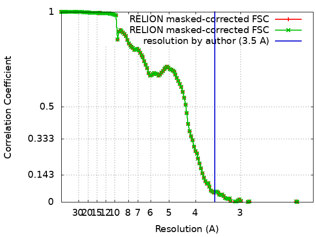 |
