 Movie
Movie Controller

[English] 日本語
 Yorodumi
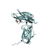
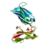
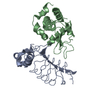
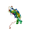
- PDB-6v3p: The BIgI domain of beta protein from S. agalactiae bound to CEACAM1 -
+ Open data

- Basic information
| Entry | Database: PDB / ID: 6v3p | ||||||
|---|---|---|---|---|---|---|---|
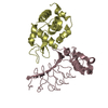
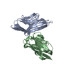
| Title | The BIgI domain of beta protein from S. agalactiae bound to CEACAM1 | ||||||
 Components Components |
| ||||||
 Keywords Keywords |  CELL ADHESION / Bacterial / Adhesin CELL ADHESION / Bacterial / Adhesin | ||||||
| Function / homology |  Function and homology information Function and homology informationregulation of endothelial cell differentiation / insulin receptor internalization / negative regulation of cytotoxic T cell degranulation / granulocyte colony-stimulating factor signaling pathway / regulation of homophilic cell adhesion / regulation of epidermal growth factor receptor signaling pathway / regulation of sprouting angiogenesis / regulation of blood vessel remodeling / negative regulation of hepatocyte proliferation / negative regulation of natural killer cell mediated cytotoxicity directed against tumor cell target ...regulation of endothelial cell differentiation / insulin receptor internalization / negative regulation of cytotoxic T cell degranulation / granulocyte colony-stimulating factor signaling pathway / regulation of homophilic cell adhesion / regulation of epidermal growth factor receptor signaling pathway / regulation of sprouting angiogenesis / regulation of blood vessel remodeling / negative regulation of hepatocyte proliferation / negative regulation of natural killer cell mediated cytotoxicity directed against tumor cell target / negative regulation of lipid biosynthetic process / bile acid transmembrane transporter activity / negative regulation of T cell mediated cytotoxicity / regulation of endothelial cell migration / filamin binding / negative regulation of granulocyte differentiation / Fibronectin matrix formation / insulin catabolic process / common myeloid progenitor cell proliferation / negative regulation of interleukin-1 production / negative regulation of fatty acid biosynthetic process / positive regulation of vasculogenesis / cell-cell adhesion via plasma-membrane adhesion molecules / regulation of immune system process / negative regulation of platelet aggregation / bile acid and bile salt transport / negative regulation of vascular permeability / wound healing, spreading of cells / microvillus membrane / transport vesicle membrane / negative regulation of T cell receptor signaling pathway / regulation of phosphatidylinositol 3-kinase/protein kinase B signal transduction / blood vessel development / homophilic cell adhesion via plasma membrane adhesion molecules / tertiary granule membrane / lateral plasma membrane / specific granule membrane / regulation of cell migration / protein tyrosine kinase binding / basal plasma membrane / regulation of ERK1 and ERK2 cascade / integrin-mediated signaling pathway / regulation of cell growth / Cell surface interactions at the vascular wall / adherens junction / negative regulation of protein kinase activity / kinase binding / cellular response to insulin stimulus / cell migration / cell-cell junction / cell junction / actin binding / protein phosphatase binding / angiogenesis / protein dimerization activity / calmodulin binding / cell adhesion / apical plasma membrane / Neutrophil degranulation / cell surface / signal transduction / protein homodimerization activity / extracellular exosome / extracellular region / membrane / identical protein binding / plasma membraneSimilarity search - Function | ||||||
| Biological species |  Homo sapiens (human) Homo sapiens (human) Streptococcus agalactiae (bacteria) Streptococcus agalactiae (bacteria) | ||||||
| Method | X-RAY DIFFRACTION / SYNCHROTRON / MOLECULAR REPLACEMENT / molecular replacement / Resolution: 3.25 Å | ||||||
 Authors Authors | Bonsor, D.A. / McCarthy, A.J. | ||||||
 Citation Citation | Journal: Embo J. / Year: 2021 Title: Bacterial protein domains with a novel Ig-like fold target human CEACAM receptors. Authors: van Sorge, N.M. / Bonsor, D.A. / Deng, L. / Lindahl, E. / Schmitt, V. / Lyndin, M. / Schmidt, A. / Nilsson, O.R. / Brizuela, J. / Boero, E. / Sundberg, E.J. / van Strijp, J.A.G. / Doran, K.S. ...Authors: van Sorge, N.M. / Bonsor, D.A. / Deng, L. / Lindahl, E. / Schmitt, V. / Lyndin, M. / Schmidt, A. / Nilsson, O.R. / Brizuela, J. / Boero, E. / Sundberg, E.J. / van Strijp, J.A.G. / Doran, K.S. / Singer, B.B. / Lindahl, G. / McCarthy, A.J. | ||||||
| History |
|
- Structure visualization
| Structure viewer | Molecule: MolmilJmol/JSmol |
|---|
- Downloads & links
-Download
| PDBx/mmCIF format | 6v3p.cif.gz | 99 KB | Display | PDBx/mmCIF format |
|---|---|---|---|---|
| PDB format | pdb6v3p.ent.gz | 74.8 KB | Display | PDB format |
| PDBx/mmJSON format | 6v3p.json.gz | Tree view | PDBx/mmJSON format | |
| Others |  Other downloads Other downloads |
-Validation report
| Arichive directory | https://data.pdbj.org/pub/pdb/validation_reports/v3/6v3pftp://data.pdbj.org/pub/pdb/validation_reports/v3/6v3p | HTTPS FTP |
|---|
-Related structure data
| Related structure data |  2gk2S S: Starting model for refinement |
|---|---|
| Similar structure data |


-Links
 PDBj
PDBj


- Assembly
| Deposited unit | 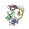
| ||||||||||||||||||||||||||||||||||||||||||||||||||||||||||||||||||||
|---|---|---|---|---|---|---|---|---|---|---|---|---|---|---|---|---|---|---|---|---|---|---|---|---|---|---|---|---|---|---|---|---|---|---|---|---|---|---|---|---|---|---|---|---|---|---|---|---|---|---|---|---|---|---|---|---|---|---|---|---|---|---|---|---|---|---|---|---|---|
| 1 | 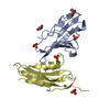
| ||||||||||||||||||||||||||||||||||||||||||||||||||||||||||||||||||||
| 2 | 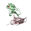
| ||||||||||||||||||||||||||||||||||||||||||||||||||||||||||||||||||||
| Unit cell |
| ||||||||||||||||||||||||||||||||||||||||||||||||||||||||||||||||||||
| Noncrystallographic symmetry (NCS) | NCS domain:
NCS domain segments: Component-ID: 0 / Refine code: 0
NCS ensembles :
|
-Components
| #1: Protein | Mass: 12101.444 Da / Num. of mol.: 2 Source method: isolated from a genetically manipulated source Source: (gene. exp.) Homo sapiens (human) / Gene: CEACAM1, BGP, BGP1 / Production host: Escherichia coli BL21(DE3) (bacteria) / References: UniProt: P13688#2: Protein | Mass: 14188.619 Da / Num. of mol.: 2 Source method: isolated from a genetically manipulated source Source: (gene. exp.) Streptococcus agalactiae (bacteria) / Gene: bag / Production host: Escherichia coli BL21(DE3) (bacteria) / References: UniProt: P27951#3: Chemical | ChemComp-SO4 / Sulfate  Mass: 96.063 Da / Num. of mol.: 5 / Source method: obtained synthetically / Formula: SO4 Mass: 96.063 Da / Num. of mol.: 5 / Source method: obtained synthetically / Formula: SO4#4: Chemical | Glycerol  Mass: 92.094 Da / Num. of mol.: 2 / Source method: obtained synthetically / Formula: C3H8O3 Mass: 92.094 Da / Num. of mol.: 2 / Source method: obtained synthetically / Formula: C3H8O3#5: Water | ChemComp-HOH / | Water Mass: 18.015 Da / Num. of mol.: 1 / Source method: isolated from a natural source / Formula: H2O Mass: 18.015 Da / Num. of mol.: 1 / Source method: isolated from a natural source / Formula: H2OHas ligand of interest | N | |
|---|
-Experimental details
-Experiment
| Experiment | Method: X-RAY DIFFRACTION / Number of used crystals: 1 |
|---|
- Sample preparation
| Crystal | Density Matthews: 5.3 Å3/Da / Density % sol: 76.77 % |
|---|---|
| Crystal grow | Temperature: 293 K / Method: vapor diffusion, hanging drop / pH: 5.5 / Details: 1.7M Ammonium Sulfate, 0.1M Sodium Citrate, pH 5.5 |
-Data collection
| Diffraction | Mean temperature: 100 K / Serial crystal experiment: N |
|---|---|
| Diffraction source | Source: SYNCHROTRON / Site: SSRL  / Beamline: BL12-2 / Wavelength: 0.9795 Å / Beamline: BL12-2 / Wavelength: 0.9795 Å |
| Detector | Type: DECTRIS PILATUS 6M / Detector: PIXEL / Date: Nov 10, 2018 Details: Mirror: Flat Si Rh coated M0, Kirkpatrick-Baez flat bent Si M1 & M |
| Radiation | Monochromator: Si (111) / Protocol: SINGLE WAVELENGTH / Monochromatic (M) / Laue (L): M / Scattering type: x-ray |
| Radiation wavelength | Wavelength: 0.9795 Å / Relative weight: 1 |
| Reflection | Resolution: 3.25→38.92 Å / Num. obs: 18198 / % possible obs: 99.8 % / Redundancy: 10.7 % / CC1/2: 0.999 / Rmerge(I) obs: 0.078 / Rpim(I) all: 0.025 / Rrim(I) all: 0.082 / Net I/σ(I): 18.2 / Num. measured all: 194550 / Scaling rejects: 13 |
| Reflection shell | Resolution: 3.25→3.51 Å / Redundancy: 10.9 % / Rmerge(I) obs: 1.411 / Num. unique obs: 3672 / CC1/2: 0.919 / Rpim(I) all: 0.445 / Rrim(I) all: 1.481 / % possible all: 99.8 |
-Phasing
| Phasing | Method: molecular replacement |
|---|
- Processing
| Software |
| ||||||||||||||||||||||||||||||||||||||||||||||||||||||||||||
|---|---|---|---|---|---|---|---|---|---|---|---|---|---|---|---|---|---|---|---|---|---|---|---|---|---|---|---|---|---|---|---|---|---|---|---|---|---|---|---|---|---|---|---|---|---|---|---|---|---|---|---|---|---|---|---|---|---|---|---|---|---|
| Refinement | Method to determine structure: MOLECULAR REPLACEMENT Starting model: 2GK2 Resolution: 3.25→38.95 Å / Cor.coef. Fo:Fc: 0.954 / Cor.coef. Fo:Fc free: 0.95 / SU B: 24.846 / SU ML: 0.354 / Cross valid method: THROUGHOUT / σ(F): 0 / ESU R: 0.675 / ESU R Free: 0.356 / Stereochemistry target values: MAXIMUM LIKELIHOOD Details: HYDROGENS HAVE BEEN ADDED IN THE RIDING POSITIONS U VALUES : REFINED INDIVIDUALLY
| ||||||||||||||||||||||||||||||||||||||||||||||||||||||||||||
| Solvent computation | Ion probe radii: 0.7 Å / Shrinkage radii: 0.7 Å / VDW probe radii: 1 Å / Solvent model: MASK | ||||||||||||||||||||||||||||||||||||||||||||||||||||||||||||
| Displacement parameters | Biso max: 305.65 Å2 / Biso mean: 149.92 Å2 / Biso min: 110.14 Å2
| ||||||||||||||||||||||||||||||||||||||||||||||||||||||||||||
| Refinement step | Cycle: final / Resolution: 3.25→38.95 Å
| ||||||||||||||||||||||||||||||||||||||||||||||||||||||||||||
| Refine LS restraints |
| ||||||||||||||||||||||||||||||||||||||||||||||||||||||||||||
| Refine LS restraints NCS | Refine-ID: X-RAY DIFFRACTION / Type: interatomic distance / Weight position: 0.05
| ||||||||||||||||||||||||||||||||||||||||||||||||||||||||||||
| LS refinement shell | Resolution: 3.25→3.334 Å / Rfactor Rfree error: 0 / Total num. of bins used: 20
|
