 Movie
Movie Controller

[English] 日本語
 Yorodumi
- PDB-1m4k: Crystal structure of the human natural killer cell activator rece... -
+ Open data

- Basic information
| Entry | Database: PDB / ID: 1m4k | ||||||
|---|---|---|---|---|---|---|---|
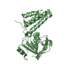
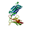
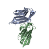
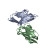
| Title | Crystal structure of the human natural killer cell activator receptor KIR2DS2 (CD158j) | ||||||
 Components Components | KILLER CELL IMMUNOGLOBULIN-LIKE RECEPTOR 2DS2 | ||||||
 Keywords Keywords |  IMMUNE SYSTEM / beta-sandwich / Ig-like C2 type domain IMMUNE SYSTEM / beta-sandwich / Ig-like C2 type domain | ||||||
| Function / homology |  Function and homology information Function and homology informationstimulatory killer cell immunoglobulin-like receptor signaling pathway / DAP12 interactions / transmembrane signaling receptor activity / Immunoregulatory interactions between a Lymphoid and a non-Lymphoid cell / immune response / membrane / plasma membraneSimilarity search - Function | ||||||
| Biological species |  Homo sapiens (human) Homo sapiens (human) | ||||||
| Method | X-RAY DIFFRACTION / SYNCHROTRON / MOLECULAR REPLACEMENT / Resolution: 2.3 Å | ||||||
 Authors Authors | Saulquin, X. / Gastinel, L.N. / Vivier, E. | ||||||
 Citation Citation | Journal: J.EXP.MED. / Year: 2003 Title: Crystal structure of the human natural killer cell activating receptor KIR2DS2 (CD158j) Authors: Saulquin, X. / Gastinel, L.N. / Vivier, E. | ||||||
| History |
|
- Structure visualization
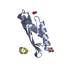
| Structure viewer | Molecule: MolmilJmol/JSmol |
|---|
- Downloads & links
-Download
| PDBx/mmCIF format | 1m4k.cif.gz | 51.3 KB | Display | PDBx/mmCIF format |
|---|---|---|---|---|
| PDB format | pdb1m4k.ent.gz | 36.6 KB | Display | PDB format |
| PDBx/mmJSON format | 1m4k.json.gz | Tree view | PDBx/mmJSON format | |
| Others |  Other downloads Other downloads |
-Validation report
| Arichive directory | https://data.pdbj.org/pub/pdb/validation_reports/m4/1m4kftp://data.pdbj.org/pub/pdb/validation_reports/m4/1m4k | HTTPS FTP |
|---|
-Related structure data
| Similar structure data |
|---|


-Links
 PDBj
PDBj


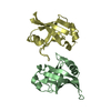
- Assembly
| Deposited unit | 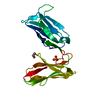
| ||||||||
|---|---|---|---|---|---|---|---|---|---|
| 1 |
| ||||||||
| Unit cell |
|
-Components
| #1: Protein | Mass: 22048.598 Da / Num. of mol.: 1 Source method: isolated from a genetically manipulated source Source: (gene. exp.) Homo sapiens (human) / Production host:  Escherichia coli (E. coli) / References: UniProt: P43631 Escherichia coli (E. coli) / References: UniProt: P43631 |
|---|---|
| #2: Chemical | ChemComp-SO4 / Sulfate  Mass: 96.063 Da / Num. of mol.: 1 / Source method: obtained synthetically / Formula: SO4 Mass: 96.063 Da / Num. of mol.: 1 / Source method: obtained synthetically / Formula: SO4 |
| #3: Chemical | ChemComp-EDO / Ethylene glycol  Mass: 62.068 Da / Num. of mol.: 1 / Source method: obtained synthetically / Formula: C2H6O2 Mass: 62.068 Da / Num. of mol.: 1 / Source method: obtained synthetically / Formula: C2H6O2 |
| #4: Water | ChemComp-HOH / Water Mass: 18.015 Da / Num. of mol.: 78 / Source method: isolated from a natural source / Formula: H2O Mass: 18.015 Da / Num. of mol.: 78 / Source method: isolated from a natural source / Formula: H2O |
-Experimental details
-Experiment
| Experiment | Method: X-RAY DIFFRACTION / Number of used crystals: 1 |
|---|
- Sample preparation
| Crystal | Density Matthews: 3.38 Å3/Da / Density % sol: 63.65 % | |||||||||||||||||||||||||||||||||||||||||||||||||
|---|---|---|---|---|---|---|---|---|---|---|---|---|---|---|---|---|---|---|---|---|---|---|---|---|---|---|---|---|---|---|---|---|---|---|---|---|---|---|---|---|---|---|---|---|---|---|---|---|---|---|
| Crystal grow | Temperature: 293 K / Method: vapor diffusion, hanging drop / pH: 4.6 Details: Ammonium sulfate, sodium acetate, Zinc chloride, pH 4.6, VAPOR DIFFUSION, HANGING DROP, temperature 293K | |||||||||||||||||||||||||||||||||||||||||||||||||
| Crystal grow | *PLUS Temperature: 20 ℃ / pH: 8 | |||||||||||||||||||||||||||||||||||||||||||||||||
| Components of the solutions | *PLUS
|
-Data collection
| Diffraction | Mean temperature: 100 K |
|---|---|
| Diffraction source | Source: SYNCHROTRON / Site: ESRF  / Beamline: ID14-4 / Wavelength: 0.9537 Å / Beamline: ID14-4 / Wavelength: 0.9537 Å |
| Detector | Type: ADSC QUANTUM 4 / Detector: CCD / Date: Apr 26, 2002 |
| Radiation | Protocol: SINGLE WAVELENGTH / Monochromatic (M) / Laue (L): M / Scattering type: x-ray |
| Radiation wavelength | Wavelength: 0.9537 Å / Relative weight: 1 |
| Reflection | Resolution: 2.1→30 Å / Num. all: 17624 / Num. obs: 17624 / % possible obs: 99.6 % / Observed criterion σ(F): 0 / Observed criterion σ(I): 0 / Redundancy: 5.1 % / Biso Wilson estimate: 31.9 Å2 / Rsym value: 0.78 / Net I/σ(I): 4.6 |
| Reflection shell | Resolution: 2.1→2.3 Å / Redundancy: 4.8 % / Mean I/σ(I) obs: 2.6 / Rsym value: 0.286 / % possible all: 99.6 |
| Reflection | *PLUS % possible obs: 61 % / Rmerge(I) obs: 0.48 |
- Processing
| Software |
| |||||||||||||||||||||
|---|---|---|---|---|---|---|---|---|---|---|---|---|---|---|---|---|---|---|---|---|---|---|
| Refinement | Method to determine structure: MOLECULAR REPLACEMENT Starting model: KIR2dL2, KIR2DL1, KIR2DL3 Resolution: 2.3→28.16 Å / σ(F): 0 / Stereochemistry target values: Engh & Huber
| |||||||||||||||||||||
| Displacement parameters |
| |||||||||||||||||||||
| Refine analyze |
| |||||||||||||||||||||
| Refinement step | Cycle: LAST / Resolution: 2.3→28.16 Å
| |||||||||||||||||||||
| Refine LS restraints |
| |||||||||||||||||||||
| LS refinement shell | Resolution: 2.3→2.44 Å / Rfactor Rfree error: 0.021
| |||||||||||||||||||||
| Refinement | *PLUS Lowest resolution: 28 Å | |||||||||||||||||||||
| Solvent computation | *PLUS | |||||||||||||||||||||
| Displacement parameters | *PLUS | |||||||||||||||||||||
| Refine LS restraints | *PLUS
|
