positive regulation of peptidase activity / myoblast differentiation / Activation of PPARGC1A (PGC-1alpha) by phosphorylation / muscle organ development / DSCAM interactions / positive regulation of muscle cell differentiation / Myogenesis / negative regulation of cell cycle / MAP kinase activity / mitogen-activated protein kinase ...positive regulation of peptidase activity / myoblast differentiation / Activation of PPARGC1A (PGC-1alpha) by phosphorylation / muscle organ development / DSCAM interactions / positive regulation of muscle cell differentiation / Myogenesis / negative regulation of cell cycle / MAP kinase activity / mitogen-activated protein kinase / signal transduction in response to DNA damage / p38MAPK events / NOD1/2 Signaling Pathway / VEGFA-VEGFR2 Pathway / Negative regulation of MAPK pathway / MAPK cascade / peptidyl-serine phosphorylation / regulation of cell cycle / intracellular signal transduction / cell cycle / protein serine kinase activity / protein serine/threonine kinase activity / magnesium ion binding / signal transduction / mitochondrion / nucleoplasm / ATP binding / nucleus / cytosol / cytoplasm Similarity search - Function
Mitogen-activated protein kinase 12 / Mitogen-activated protein (MAP) kinase p38-like / Mitogen-activated protein (MAP) kinase, conserved site / MAP kinase signature. / Protein kinase domain / Serine/Threonine protein kinases, catalytic domain / Protein kinase, ATP binding site / Protein kinases ATP-binding region signature. / Protein kinase domain profile. / Protein kinase domain / Protein kinase-like domain superfamily Similarity search - Domain/homology
 Movie
Movie Controller


 Open data
 Basic information
 Components
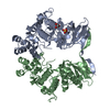
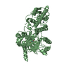
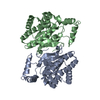
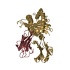
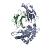
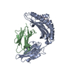
 P38 mitogen-activated protein kinases
  Keywords
 Function and homology information
 Authors
 United States, 4items
  Citation
 Structure visualization
 Downloads & links
 Other downloads


 PDBj
PDBj


 Assembly