 ムービー
ムービー コントローラー


+ データを開く

- 基本情報
基本情報
| 登録情報 | データベース: PDB / ID: 6ufr | ||||||||||||
|---|---|---|---|---|---|---|---|---|---|---|---|---|---|
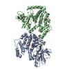
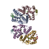
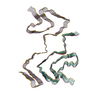
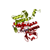
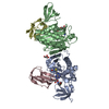
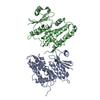
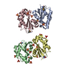
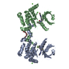
| タイトル | Structure of recombinantly assembled E46K alpha-synuclein fibrils | ||||||||||||
 要素 要素 | Alpha-synuclein Α-シヌクレイン Α-シヌクレイン | ||||||||||||
 キーワード キーワード | PROTEIN FIBRIL / alpha-synuclein (Α-シヌクレイン) / amyloid (アミロイド) / fibril (フィブリル) / E46K / hereditary mutations / parkinson's disease (パーキンソン病) / lewy body dementia | ||||||||||||
| 機能・相同性 |  機能・相同性情報 機能・相同性情報negative regulation of mitochondrial electron transport, NADH to ubiquinone / neutral lipid metabolic process / regulation of phospholipase activity / negative regulation of monooxygenase activity / regulation of acyl-CoA biosynthetic process / negative regulation of dopamine uptake involved in synaptic transmission / negative regulation of norepinephrine uptake / positive regulation of glutathione peroxidase activity / positive regulation of SNARE complex assembly / positive regulation of hydrogen peroxide catabolic process ...negative regulation of mitochondrial electron transport, NADH to ubiquinone / neutral lipid metabolic process / regulation of phospholipase activity / negative regulation of monooxygenase activity / regulation of acyl-CoA biosynthetic process / negative regulation of dopamine uptake involved in synaptic transmission / negative regulation of norepinephrine uptake / positive regulation of glutathione peroxidase activity / positive regulation of SNARE complex assembly / positive regulation of hydrogen peroxide catabolic process / supramolecular fiber / negative regulation of transporter activity / mitochondrial membrane organization / negative regulation of chaperone-mediated autophagy / regulation of reactive oxygen species biosynthetic process / positive regulation of protein localization to cell periphery / regulation of synaptic vesicle recycling / negative regulation of platelet-derived growth factor receptor signaling pathway / negative regulation of exocytosis / regulation of glutamate secretion / response to iron(II) ion / regulation of norepinephrine uptake / dopamine biosynthetic process / SNARE complex assembly / positive regulation of neurotransmitter secretion / regulation of locomotion / positive regulation of inositol phosphate biosynthetic process / synaptic vesicle priming / regulation of macrophage activation / dopamine uptake involved in synaptic transmission / negative regulation of microtubule polymerization / synaptic vesicle transport / dynein complex binding / positive regulation of receptor recycling / regulation of dopamine secretion / protein kinase inhibitor activity / negative regulation of thrombin-activated receptor signaling pathway / response to type II interferon / cuprous ion binding / positive regulation of exocytosis / synaptic vesicle exocytosis / mitochondrial ATP synthesis coupled electron transport / positive regulation of endocytosis / kinesin binding / cysteine-type endopeptidase inhibitor activity involved in apoptotic process / response to magnesium ion / synaptic vesicle endocytosis / regulation of presynapse assembly / alpha-tubulin binding / negative regulation of serotonin uptake / localization / phospholipid metabolic process / axon terminus / supramolecular fiber organization / 封入体 / cellular response to copper ion / Hsp70 protein binding / cellular response to epinephrine stimulus / excitatory postsynaptic potential / response to interleukin-1 / adult locomotory behavior / SNARE binding / positive regulation of release of sequestered calcium ion into cytosol / fatty acid metabolic process / long-term synaptic potentiation / regulation of transmembrane transporter activity / phosphoprotein binding / protein tetramerization / synapse organization / regulation of long-term neuronal synaptic plasticity / microglial cell activation / negative regulation of protein kinase activity / ferrous iron binding / protein destabilization / PKR-mediated signaling / negative regulation of cysteine-type endopeptidase activity involved in apoptotic process / tau protein binding / positive regulation of protein serine/threonine kinase activity / receptor internalization / phospholipid binding / synaptic vesicle membrane / positive regulation of inflammatory response / activation of cysteine-type endopeptidase activity involved in apoptotic process / マイクロフィラメント / positive regulation of peptidyl-serine phosphorylation / actin binding / cellular response to oxidative stress / 細胞皮質 / histone binding / 成長円錐 / postsynapse / chemical synaptic transmission / neuron apoptotic process / negative regulation of neuron apoptotic process / response to lipopolysaccharide / amyloid fibril formation / リソソーム / molecular adaptor activity / oxidoreductase activity / transcription cis-regulatory region binding類似検索 - 分子機能 | ||||||||||||
| 生物種 |  Homo sapiens (ヒト) Homo sapiens (ヒト) | ||||||||||||
| 手法 | 電子顕微鏡法 / らせん対称体再構成法 / クライオ電子顕微鏡法 / 解像度: 2.5 Å | ||||||||||||
 データ登録者 データ登録者 | Eisenberg, D.S. / Boyer, D.R. / Sawaya, M.R. / Li, B. / Jiang, L. | ||||||||||||
| 資金援助 |  米国, 3件 米国, 3件
| ||||||||||||
 引用 引用 | ジャーナル: Proc Natl Acad Sci U S A / 年: 2020 タイトル: The α-synuclein hereditary mutation E46K unlocks a more stable, pathogenic fibril structure. 著者: David R Boyer / Binsen Li / Chuanqi Sun / Weijia Fan / Kang Zhou / Michael P Hughes / Michael R Sawaya / Lin Jiang / David S Eisenberg / 要旨: Aggregation of α-synuclein is a defining molecular feature of Parkinson's disease, Lewy body dementia, and multiple systems atrophy. Hereditary mutations in α-synuclein are linked to both ...Aggregation of α-synuclein is a defining molecular feature of Parkinson's disease, Lewy body dementia, and multiple systems atrophy. Hereditary mutations in α-synuclein are linked to both Parkinson's disease and Lewy body dementia; in particular, patients bearing the E46K disease mutation manifest a clinical picture of parkinsonism and Lewy body dementia, and E46K creates more pathogenic fibrils in vitro. Understanding the effect of these hereditary mutations on α-synuclein fibril structure is fundamental to α-synuclein biology. We therefore determined the cryo-electron microscopy (cryo-EM) structure of α-synuclein fibrils containing the hereditary E46K mutation. The 2.5-Å structure reveals a symmetric double protofilament in which the molecules adopt a vastly rearranged, lower energy fold compared to wild-type fibrils. We propose that the E46K misfolding pathway avoids electrostatic repulsion between K46 and K80, a residue pair which form the E46-K80 salt bridge in the wild-type fibril structure. We hypothesize that, under our conditions, the wild-type fold does not reach this deeper energy well of the E46K fold because the E46-K80 salt bridge diverts α-synuclein into a kinetic trap-a shallower, more accessible energy minimum. The E46K mutation apparently unlocks a more stable and pathogenic fibril structure. | ||||||||||||
| 履歴 |
|
- 構造の表示
構造の表示
| ムービー |
ムービービューア |
|---|---|
| 構造ビューア | 分子: MolmilJmol/JSmol |
- ダウンロードとリンク
-ダウンロード
| PDBx/mmCIF形式 | 6ufr.cif.gz | 113.8 KB | 表示 | PDBx/mmCIF形式 |
|---|---|---|---|---|
| PDB形式 | pdb6ufr.ent.gz | 84.7 KB | 表示 | PDB形式 |
| PDBx/mmJSON形式 | 6ufr.json.gz | ツリー表示 | PDBx/mmJSON形式 | |
| その他 |  その他のダウンロード その他のダウンロード |
-検証レポート
| アーカイブディレクトリ | https://data.pdbj.org/pub/pdb/validation_reports/uf/6ufrftp://data.pdbj.org/pub/pdb/validation_reports/uf/6ufr | HTTPS FTP |
|---|
-関連構造データ
| 関連構造データ | 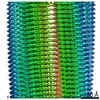 20759MC M: このデータのモデリングに利用したマップデータ C: 同じ文献を引用 ( |
|---|---|
| 類似構造データ | |
| 電子顕微鏡画像生データ | EMPIAR-10494 (タイトル: The α-synuclein hereditary mutation E46K unlocks a more stable, pathogenic fibril structure Data size: 897.9 Data #1: Unaligned K3 frames from alpha-synuclein E46K amyloid fibrils [micrographs - multiframe]) |
-リンク
 PDBj
PDBj
- 集合体
集合体
| 登録構造単位 | 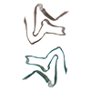
|
|---|---|
| 1 |
|
-要素
| #1: タンパク質 | Α-シヌクレイン / Non-A beta component of AD amyloid / Non-A4 component of amyloid precursor / NACP 分子量: 14476.175 Da / 分子数: 10 / 変異: E46K / 由来タイプ: 組換発現 / 由来: (組換発現) Homo sapiens (ヒト) / 遺伝子: SNCA, NACP, PARK1 / 発現宿主:  Escherichia coli (大腸菌) / 参照: UniProt: P37840 Escherichia coli (大腸菌) / 参照: UniProt: P37840#2: 水 | ChemComp-HOH / | 水 分子量: 18.015 Da / 分子数: 90 / 由来タイプ: 天然 / 式: H2O 分子量: 18.015 Da / 分子数: 90 / 由来タイプ: 天然 / 式: H2O |
|---|
-実験情報
-実験
| 実験 | 手法: 電子顕微鏡法 |
|---|---|
| EM実験 | 試料の集合状態: FILAMENT / 3次元再構成法: らせん対称体再構成法 |
- 試料調製
試料調製
| 構成要素 | 名称: Recombinantly assembled E46K alpha-synuclein fibril / タイプ: ORGANELLE OR CELLULAR COMPONENT / Entity ID: #1 / 由来: RECOMBINANT |
|---|---|
| 由来(天然) | 生物種: Homo sapiens (ヒト) |
| 由来(組換発現) | 生物種: Escherichia coli (大腸菌) |
| 緩衝液 | pH: 7 |
| 試料 | 包埋: NO / シャドウイング: NO / 染色: NO / 凍結: YES |
| 急速凍結 | 凍結剤: ETHANE |
- 電子顕微鏡撮影
| 実験機器 |  モデル: Titan Krios / 画像提供: FEI Company |
|---|---|
| 顕微鏡 | モデル: FEI TITAN KRIOS |
| 電子銃 | 電子線源: FIELD EMISSION GUN / 加速電圧: 300 kV / 照射モード: FLOOD BEAM |
| 電子レンズ | モード: BRIGHT FIELDBright-field microscopy |
| 撮影 | 電子線照射量: 36 e/Å2 / 検出モード: SUPER-RESOLUTION フィルム・検出器のモデル: GATAN K2 SUMMIT (4k x 4k) |
- 解析
解析
| ソフトウェア | 名称: PHENIX / バージョン: 1.16_3549: / 分類: 精密化 | ||||||||||||||||||||||||
|---|---|---|---|---|---|---|---|---|---|---|---|---|---|---|---|---|---|---|---|---|---|---|---|---|---|
| CTF補正 | タイプ: PHASE FLIPPING AND AMPLITUDE CORRECTION | ||||||||||||||||||||||||
| らせん対称 | 回転角度/サブユニット: 178.92 ° / 軸方向距離/サブユニット: 4.85 Å / らせん対称軸の対称性: C2 | ||||||||||||||||||||||||
| 3次元再構成 | 解像度: 2.5 Å / 解像度の算出法: FSC 0.143 CUT-OFF / 粒子像の数: 114260 / 対称性のタイプ: HELICAL | ||||||||||||||||||||||||
| 拘束条件 |
|
