 Movie
Movie Controller

[English] 日本語
 Yorodumi
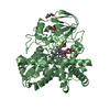
- PDB-2ejf: Crystal Structure Of The Biotin Protein Ligase (Mutations R48A an... -
+ Open data

- Basic information
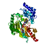
| Entry | Database: PDB / ID: 2ejf | ||||||
|---|---|---|---|---|---|---|---|
| Title | Crystal Structure Of The Biotin Protein Ligase (Mutations R48A and K111A) and Biotin Carboxyl Carrier Protein Complex From Pyrococcus Horikoshii OT3 | ||||||
 Components Components |
| ||||||
 Keywords Keywords |  LIGASE / Biotinylation / Dimer / Structural Genomics / NPPSFA / National Project on Protein Structural and Functional Analyses / RIKEN Structural Genomics/Proteomics Initiative / RSGI LIGASE / Biotinylation / Dimer / Structural Genomics / NPPSFA / National Project on Protein Structural and Functional Analyses / RIKEN Structural Genomics/Proteomics Initiative / RSGI | ||||||
| Function / homology |  Function and homology information Function and homology informationbiotin-[acetyl-CoA-carboxylase] ligase activity / protein modification process / ATP bindingSimilarity search - Function | ||||||
| Biological species |   Pyrococcus horikoshii (archaea) Pyrococcus horikoshii (archaea) | ||||||
| Method | X-RAY DIFFRACTION / SYNCHROTRON / MOLECULAR REPLACEMENT / Resolution: 2 Å | ||||||
 Authors Authors | Bagautdinov, B. / Matsuura, Y. / Bagautdinova, S. / Kunishima, N. / RIKEN Structural Genomics/Proteomics Initiative (RSGI) | ||||||
 Citation Citation | Journal: J.Biol.Chem. / Year: 2008 Title: Protein biotinylation visualized by a complex structure of biotin protein ligase with a substrate Authors: Bagautdinov, B. / Matsuura, Y. / Bagautdinova, S. / Kunishima, N. | ||||||
| History |
|
- Structure visualization
| Structure viewer | Molecule: MolmilJmol/JSmol |
|---|
- Downloads & links
-Download
| PDBx/mmCIF format | 2ejf.cif.gz | 140.7 KB | Display | PDBx/mmCIF format |
|---|---|---|---|---|
| PDB format | pdb2ejf.ent.gz | 107.4 KB | Display | PDB format |
| PDBx/mmJSON format | 2ejf.json.gz | Tree view | PDBx/mmJSON format | |
| Others |  Other downloads Other downloads |
-Validation report
| Arichive directory | https://data.pdbj.org/pub/pdb/validation_reports/ej/2ejfftp://data.pdbj.org/pub/pdb/validation_reports/ej/2ejf | HTTPS FTP |
|---|
-Related structure data
| Related structure data |  1x01C  2d5dSC 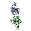 2dxuC  2dzcC  2e41C  2e64SC  2ejgC 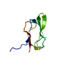 2evbC  2zgwC C: citing same article ( S: Starting model for refinement |
|---|---|
| Similar structure data | |
| Other databases |


-Links
 PDBj
PDBj


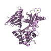
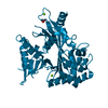
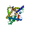
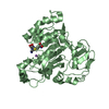
- Assembly
| Deposited unit | 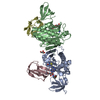
| ||||||||
|---|---|---|---|---|---|---|---|---|---|
| 1 | 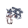
| ||||||||
| 2 | 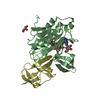
| ||||||||
| Unit cell |
|
-Components
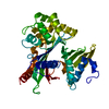
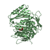
-Protein , 2 types, 4 molecules ABCD
| #1: Protein | Mass: 25959.305 Da / Num. of mol.: 2 / Mutation: R48A/K111A Source method: isolated from a genetically manipulated source Source: (gene. exp.) Pyrococcus horikoshii (archaea) / Strain: OT3 / Gene: bpl / Plasmid: pET11a / Production host:  Escherichia coli (E. coli) / Strain (production host): (DE3)RIL / References: UniProt: O57883 Escherichia coli (E. coli) / Strain (production host): (DE3)RIL / References: UniProt: O57883#2: Protein | Mass: 7985.457 Da / Num. of mol.: 2 / Fragment: residues 77-149 Source method: isolated from a genetically manipulated source Source: (gene. exp.) Pyrococcus horikoshii (archaea) / Strain: OT3 / Gene: bccp / Plasmid: pET11a / Production host: Escherichia coli (E. coli) / Strain (production host): (DE3)RIL / References: UniProt: O59021 |
|---|
-Non-polymers , 4 types, 415 molecules 


| #3: Chemical | Biotin Mass: 244.311 Da / Num. of mol.: 3 / Source method: obtained synthetically / Formula: C10H16N2O3S Mass: 244.311 Da / Num. of mol.: 3 / Source method: obtained synthetically / Formula: C10H16N2O3S#4: Chemical | Adenosine Mass: 267.241 Da / Num. of mol.: 2 / Source method: obtained synthetically / Formula: C10H13N5O4 Mass: 267.241 Da / Num. of mol.: 2 / Source method: obtained synthetically / Formula: C10H13N5O4#5: Chemical | ChemComp-GOL / | Glycerol Mass: 92.094 Da / Num. of mol.: 1 / Source method: obtained synthetically / Formula: C3H8O3 Mass: 92.094 Da / Num. of mol.: 1 / Source method: obtained synthetically / Formula: C3H8O3#6: Water | ChemComp-HOH / | WaterMass: 18.015 Da / Num. of mol.: 409 / Source method: isolated from a natural source / Formula: H2O |
|---|
-Experimental details
-Experiment
| Experiment | Method: X-RAY DIFFRACTION / Number of used crystals: 1 |
|---|
- Sample preparation
| Crystal | Density Matthews: 2.44 Å3/Da / Density % sol: 49.63 % |
|---|---|
| Crystal grow | Temperature: 295 K / Method: microbatch / pH: 4.96 Details: 10.5w/v(%) PEG 20000, 0.1M Acet, NaOH, pH 4.96, microbatch, temperature 295K |
-Data collection
| Diffraction | Mean temperature: 100 K |
|---|---|
| Diffraction source | Source: SYNCHROTRON / Site: SPring-8  / Beamline: BL26B1 / Wavelength: 1 Å / Beamline: BL26B1 / Wavelength: 1 Å |
| Detector | Type: MARRESEARCH / Detector: CCD / Date: Dec 12, 2006 / Details: MIRRORS |
| Radiation | Monochromator: GRAPHITE / Protocol: SINGLE WAVELENGTH / Monochromatic (M) / Laue (L): M / Scattering type: x-ray |
| Radiation wavelength | Wavelength: 1 Å / Relative weight: 1 |
| Reflection | Resolution: 2→37.62 Å / Num. obs: 42136 / % possible obs: 95.6 % / Observed criterion σ(I): 0 / Redundancy: 1.8 % / Biso Wilson estimate: 21.7 Å2 / Rmerge(I) obs: 0.087 / Rsym value: 0.071 / Net I/σ(I): 8.6 |
| Reflection shell | Resolution: 2→2.07 Å / Redundancy: 1.7 % / Rmerge(I) obs: 0.313 / Mean I/σ(I) obs: 2.3 / Num. unique all: 3935 / Rsym value: 0.298 / % possible all: 90 |
- Processing
| Software |
| ||||||||||||||||||||||||||||||||||||||||||||||||||||||||||||
|---|---|---|---|---|---|---|---|---|---|---|---|---|---|---|---|---|---|---|---|---|---|---|---|---|---|---|---|---|---|---|---|---|---|---|---|---|---|---|---|---|---|---|---|---|---|---|---|---|---|---|---|---|---|---|---|---|---|---|---|---|---|
| Refinement | Method to determine structure: MOLECULAR REPLACEMENT Starting model: PDB ENTRIES 2E64 AND 2D5D Resolution: 2→37.62 Å / Isotropic thermal model: OVERALL / Cross valid method: THROUGHOUT / σ(F): 0 / Stereochemistry target values: ENGH & HUBER
| ||||||||||||||||||||||||||||||||||||||||||||||||||||||||||||
| Displacement parameters | Biso mean: 28.7 Å2
| ||||||||||||||||||||||||||||||||||||||||||||||||||||||||||||
| Refine analyze |
| ||||||||||||||||||||||||||||||||||||||||||||||||||||||||||||
| Refinement step | Cycle: LAST / Resolution: 2→37.62 Å
| ||||||||||||||||||||||||||||||||||||||||||||||||||||||||||||
| Refine LS restraints |
|
